Research Article
Molecular Cloning and Bioinformatics Analysis of T3SS Inner Membrane Ring HrpQ from Vibrio harveyi 
2 Guangdong Provincial Key Laboratory of Pathogenic Biology and Epidemiology for Aquatic Animals, Zhanjiang, 524088, China
3 Key Laboratory of Control for Diseases of Aquatic Animals of Guangdong Higher Education Institutes, Zhanjiang, 524088, China
 Author
  Correspondence author
Genomics and Applied Biology, 2018, Vol. 9, No. 7 doi: 10.5376/gab.2018.09.0007
Received: 15 Aug., 2018 Accepted: 23 Sep., 2018 Published: 10 Sep., 2018
Kuebutornye F.K.A., Liao J.M., Pang H.Y., Lu Y.S., Ayiku S., and Sakyi M.E., 2018, Molecular cloning and bioinformatics analysis of T3SS inner membrane ring HrpQ from Vibrio harveyi, Genomics and Applied Biology, 9(7): 40-47 (doi: 10.5376/gab.2018.09.0007)
In this study, Vibrio harveyi strain HY99 was isolated from a diseased Epinephelus coioides. A full-length HrpQ gene of the bacteria was cloned and the amino acid sequence analyzed. The length of HrpQ gene sequence was 1,302 bp and coded 433 Amino acids. HrpQ relative molecular weight of theoretical prediction and isoelectric point were 48.002 kDa and 5.08 respectively. This protein was hydrophilic and there was one transmembrane region. There were three N-glycosylation sites. Structural analysis showed that the protein belonged to the FHA, Yop-YscD ppl superfamily. Secondary structure was composed of 31.41% α-helices, 21.71% extension chains, 7.16% beta turns and 39.72% random curls. The 3D structure of HrpQ protein was predicted to be a monomer similar to the 3D structure of the YscD putative type III secretion protein confirming that HrpQ is a T3SS protein and can be activated in vivo. This study provides a theoretical basis for further study on the function of HrpQ protein.
Background
Vibrio harveyi is gram-negative, ubiquitous bacteria which can be found in marine and estuarine ecosystems and usually known for the infection of larval and juvenile penaeids (Hashem and El-Barbary, 2013) and cause other opportunistic diseases in fish (Chatterjee and Haldar, 2002; Zorrilla et al., 2003). They can also be found in the gut of some marine animals (Hashem and El-Barbary, 2013). V. harveyi is linked with vibriosis in Acanthurus sohal (Arabian Surgeon) (Hashem and El-Barbary, 2013), Peneause monodon (Tiger prawn), Litopenaeus vannamei (White shrimp) Epinephelus coioides (Grouper) and Sulculus diversicolor (Japanese abalone) (Chatterjee and Haldar, 2002; Pang et al., 2016). The great economic loss in marine-based aquaculture in Asia has been associated with V. harveyi (Pang et al., 2010; Ransangan et al., 2012). Anorexia, tail and fin rot, local hemorrhagic ulcers on the mouth or skin surface, focal necrotic lesions in the muscle and swollen intestine, darkening of the whole fish and eye opacity are some of the symptoms characterized with Vibriosis caused by V. harveyi (Ransangan and Mustafa, 2009).
Researchers revealed that Type III secretion system (T3SS) is associated with Gram-negative pathogenic bacteria to deliver bacterial effector proteins into host cells and the effect is the alteration of host cellular functions. The Type III secretion system (T3SS) is a protein export pathway used by Gram-negative bacteria and delivers effector proteins directly into the eukaryotic cell (Ghosh, 2004; Portaliou et al., 2016). It is situated in the pathogenicity islands (PAIs) of bacteria plasmids or chromosomes (Li, 2016). Effector proteins are known to possess diverse functions but usually, help the pathogen in invading the host tissue, subdue its immune system, or in some cases help the pathogen to survive (Viboud et al., 2005; Mattoo et al., 2007).
The T3SS needle seems to be to be placed on a pair of concentric membrane-embedded rings, the larger concentric rings situated in the inner membrane and the smaller in the outer membrane, including the peptidoglycan layer. The inner membrane ring is the larger of the two coaxial rings, and protein components that make up the inner ring have been identified for a number of bacteria. For example in Salmonella, the inner membrane ring is formed by the proteins PrgK and PrgH, and in Shigella it is formed by the proteins MxiJ and MxiG (Ghosh, 2004; Coburn et al., 2007).
Various inner membrane proteins have been predicted and these predicted inner membrane T3SS proteins possess the ability to interact with effector proteins during transit and may act as receptors that identify secretion signals on effector proteins, for example Yersinia YscV also known as LcrD is predicted to possess a large cytoplasmic domain which may serve as a possible receptor site for interaction with translocation substrates (Ghosh, 2004). YscV is also said to play a role in effector secretion (Lavander et al., 2002). It was also demonstrated that overexpression of the cytoplasmic domain of YscU in wild-type Yersinia increases effector secretion (Lavander et al., 2002). Other predicted inner membranes proteins include YscR, YscS, YscTATPase and YscQ (Macnab, 2003; Ghosh, 2004). In this study, HrpQ gene of Vibrio harveyi HY99 was cloned, sequenced and bioinformatically analyzed to provide a theoretical basis for further study on the function of HrpQ protein.
1 Materials and Methods
1.1 Materials
1.1.1 Strain and vector
Vibrio harveyi strain HY99 which was isolated from a diseased Epinephelus coioides in Zhanjiang Harbor in Guangdong Province was preserved in College of Fisheries, Guangdong Ocean University. The cloning vector pMD18-T (Amp+) was purchased from TaKaRa Biotechnology (Dalian) Co., Ltd. Escherichia coli DH5α was preserved in our laboratory.
1.1.2 Reagents
Ampicillin stock solution (100 mg/mL) was prepared with sterile water and stored at -20°C. The working concentration was 100 µL/100 ml in the medium. Chloramphenicol stock solution was prepared with anhydrous ethanol and the working concentration was 25 µg/ml. Calcium chloride solution (0.1 mol./L) used to prepare competent cells was prepared by dissolving 1.7 g of CaCl2.2H2O in 100 ml of sterile water, filtered through 0.22 µm filter and stored in refrigerator at 4°C.
UNIQ-10 Column Bacteria Genomic DNA Extraction Kit was purchased from Sangon Biotech (Shanghai) Co., Ltd., EsayPure Plasmid MiniPrep Kit and EasyPureTM Quick Gel Extraction Kit were purchased from Beijing Transgen Biotech Co., Ltd., TIANamp Bacteria DNA Kit was purchased from Tiangen Biotech (Beijing) Co., Ltd.
TaKaRaEx taq DNA polymerase and T4 DNA ligase were purchased from TaKaRa Biotechnology (Dalian) Co., Ltd., EsayPfu DNA polymerase was purchased from Tiangen Biotech (Beijing) Co., Ltd.
Primer synthesis and sequencing were done by Sangon Biotech Co., Ltd. (Guangzhou).
The media used in this experiment include TSA medium (pH 7.3±0.2), TSB medium (pH 7.3±0.2), LB liquid medium and LB solid medium (containing 1.5% agar).
1.2 Methods
1.2.1 Extraction of genomic DNA of V. harveyi strain HY99
V. harveyi was inoculated to TSB medium at 28°C for 18 h. 1 mL of bacterial solution was transferred to a centrifuge tube and centrifuged at 5,000 r/min for 5 min. After removal of the supernatant, the cells were washed twice with PBS, The precipitate was resuspended with 200 μL of double distilled water using a pipette, boiled with 100°C water for 5 min and centrifuged at 12,000 r/min for 5 minutes. The supernatant was transferred to a sterile EP tube as genomic DNA template and preserved at -20°C before use.
1.2.2 Cloning of HrpQ gene from V. harveyi strain HY99
Two primer pairs were designed based on the full-length genome sequence of V. harveyi (GenBank accession number: KNY47298.1) using Primer ver. 5.0 software. The forward primer was 5’-TGGAAAATT CGTATCCTCTCCGGTG-3’ and the reverse primer was 5’-TCAAGATTTGTCATTGG TATTTTCAC-3’. The total DNA was taken as a template for amplification.
The PCR reaction system contained 1 µL of each primer, 1 µL of extracted DNA sample from V. harveyi HY99, 12.5 µL of 10×Extaq buffer, 9.5 µL of double distilled water.
The PCR amplification was started with initial denaturation at 96°C for 5 min followed by 34 cycles of denaturation at 96°C for 30 s, annealing at 57°C for 45 s, and extension at 72°C for 60 s; the amplification was completed by holding the reaction mixture at 72°C for 10 min. PCR products were detected using 5 µL product by 1% agarose gel electrophoresis and purified using gel recovery kit.
1.2.3 Ligation and sequencing of the target fragment
The purified PCR products were ligated with pMD18-T vector at 16°C for 10 h and transformed into E.coli DH5α competent cells. Positive colonies were identified by colony PCR and sequenced by Guangzhou Sangon Biotech Co. Ltd.
1.2.4 Bioinformatics analysis
ExPASy Proteomics Server was applied to deduce the amino acid sequences, identify the open reading frame and predict molecular weight and theoretical isoelectric point (pI). The sequence alignment was performed in the NCBI. The amino acid homology was analyzed using Gene DoC software. The transmembrane domain was predicted using TMHMM Server 2.0. The signal peptide sequence was predicted using SignalP 4.1 Server. The distribution of functional sites of amino acid sequences was analyzed using SoftBerry-Psite. Subcellular localization was predicted using PSORT II Prediction. The phylogenetic tree was constructed using Clastal 2.0 and MEGA 5.0. Functional domains were predicted using SMART. The three-dimensional structure was constructed using SWISS-MODEL Workspace (Arnold et al., 2006; Pang et al., 2014).
2 Results
2.1 Full-length HrpQ gene
After electrophoresis, 1,302 bp of the target gene was observed as expected (Figure 1). Then it was gel extracted, ligated intpMD 18-T vector and transformed into DH5α, and a DNA fragment of 1,302 bp was also amplified through colony PCR (Figure 1). The sequencing result revealed that HrpQ gene contained 1,302 bp open reading frame (ORF) encoding a putative protein of 433 amino acids. The obtained sequence was submitted to the NCBI GenBank (accession number: MH193372). Blast analysis results revealed that the amplification of gene nucleotide sequences had a very high homology with MULTISPECIES: EscD/YscD/HrpQ family type III secretion system inner membrane ring protein (Vibrio) and EscD/YscD/HrpQ family type III secretion system inner membrane ring protein (Vibrio harveyi) reported in NCBI gene sequences (GenBank Accession No.WP_005450275 and WP_050933567 respectively). The deduced amino acid sequences homologies were as high as 100% and 99% respectively.
|
Figure 1 Cloning (A) and identification (B) of HrpQ gene Note: Lane M: DNA marker DL2000; Lanes 1-4: HrpQ PCR products; Lanes 5-9: colony PCR products |
.png)
2.2 Characteristics of HrpQ protein in V. harveyi
Using SoftBerry-Psite software, it was predicted that HrpQ protein had three N-glycosylation sites, seven protein kinase C phosphorylation sites, eight casein kinase II phosphorylation sites, six N-myristoylation sites, one prenyl group binding sites and six microbodies C-terminal targeting signal (Figure 2). Logging on the website (http://web.expasy.org/protparam/) for online analysis, the formula of protein (HrpQ) was C2131H3433N585O656S8 and the protein was composed of 6,813 atoms. ExPASy analysis showed that HrpQ had a molecular mass of 48.002 kDa with a pI of 5.08 and an instability index (II) of 31.04, there were 63 negatively charged residues (Asp+Glu) and 50 positively charged residues (Arg+Lys); the grand average of hydropathicity (GRAVY) was -0.116. Online analysis also predicted that hydrophobic amino acids GRAVY were greater than zero, and the hydrophilic amino acids GRAVY were less than zero.
|
Figure 2 HrpQ gene sequence and amino acid sequence
Note: |
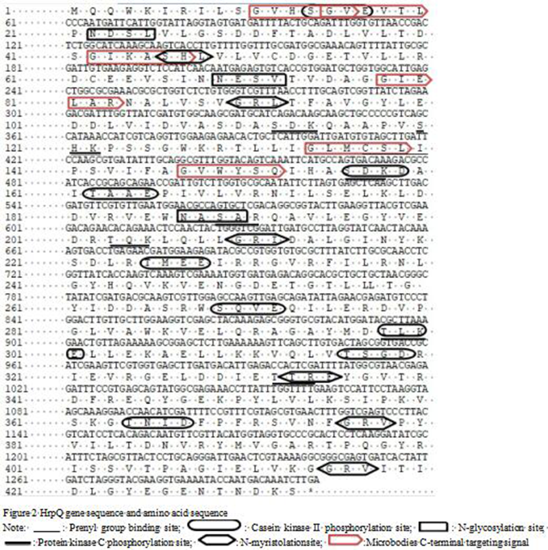
.jpg) : Prenyl group binding site;
: Prenyl group binding site; .jpg) : Casein kinase II phosphorylation site;
: Casein kinase II phosphorylation site; .jpg) : N-glycosylation site;
: N-glycosylation site; .jpg) : Protein kinase C phosphorylation site;
: Protein kinase C phosphorylation site;  : N-myristolationsite;
  : Microbodies C-terminal targeting signal
2.3 Functional domains and secondary structure of HrpQ protein
The signal peptide sequence of HrpQ protein was predicted using SignalP 4.0 Server. The results indicated that the protein had no signal peptide. The secondary structure of HrpQ protein was predicted using SOPMA software. The results suggested that HrpQ protein composed of 31.41% α-helices, 21.71% extension chains, 7.16% beta turns and 39.72% random curls (Figure 3). Using TMHMM Server 2.0 software, it was predicted that HrpQ protein contained one transmembrane domain. It was also predicted that HrpQ protein belonged to Yop-YscD_ppl superfamilies (Figure 4).
|
Figure 3 Prediction of secondary structure of HrpQ protein |
.png)
|
Figure 4 Analysis of domain of HrpQ protein |
.png)
2.4 Homology analysis with similar protein sequences of other Vibrio strains
The HrpQ protein sequence of V. harveyi was subjected to homology comparison with other Vibrios trains including V. campbelli and V. jasicida through BLAST (http://blast.ncbi.nlm.nih.Gov/Blast.cgi). As shown in Figure 5, the HrpQ gene of V.harveyi HY99 had a homology of 100% and 99% to MULTISPECIES: EscD/YscD/HrpQ family type III secretion system inner membrane ring protein (Vibrio) (accession number: WP_005450275.1) and EscD/YscD/HrpQ family type III secretion system inner membrane ring protein (Vibrio harveyi) (accession number: WP_050933567.1).
|
Figure 5 Homology comparison of amino acid sequence of Vibrio HrpQ with other bacteria |
.png)
2.5 Phylogenetic analysis
The phylogenetic tree was constructed using MEGA 5.0 software following multiple sequence alignment. As shown in Figure 6, HrpQ from V. harveyi was clustered into the same subgroup with corresponding proteins from V. campbellii and V. jasicida, indicating close genetic relationships.
|
Figure 6 Phylogenetic tree of HrpQ constructed by neighbour joining method |
.png)
2.6 Three-dimensional structure of the conserved domain of HrpQ protein
SWISS Model Server was used to build the three-dimensional (3D) structure and to predict the secondary structure of HrpQ protein (Figure 7). The 3D structure of HrpQ protein was predicted to be a monomer similar to the 3D structure of the YscD putative type III secretion protein detected by Xray diffraction. The consistency of both sequences was 31.36%. The resolution was 2.5A.
|
Figure 7 Three-dimensional structure of HrpQ protein |
.png)
3 Discussion
The ability of Gram-negative bacteria to colonize its host depends on the type III secretion (T3SS) system, which transports selected bacterial proteins into host cells (Gamez et al., 2012). The T3SS needle appears to be positioned on a pair of membranes; inner and outer and these predicted inner membrane T3SS proteins may interact with effector proteins during transit and possibly serve as receptors that recognize secretion signals on effector proteins (Ghosh, 2004). In this study, the inner membrane gene HrpQ of V. harveyi HY99 was cloned and homology comparison and phylogenetic analysis revealed that the inner membrane protein HrpQ of V. harveyi HY99 had a close homology with the HrpQ of V. campbellii and V. jasicida which satisfies the results of morphological and biochemical classification. These results suggested that the inner membrane protein HrpQ plays an important role in the pathogenesis of Vibrio.
The result revealed that HrpQ protein of V. harveyi HY99 has no signal peptide which suggested that after the synthesis of HrpQ, the protein was not transported therefore it was not a secretory protein or some cytoplasmic matrix synthesis proteins (Sui et al., 2016).
An important step of protein modification after translation is N-glycosylation. N-glycosylation influences biological behaviour of protein such as antigenicity, folding, transport, positioning and stability (Sui et al., 2016). From the results, it is evident that HrpQ of V. harveyi HY99 had three N-glycosylation sites. In addition, the HrpQ peptide chain had multiple functional regions namely protein kinase C phosphorylation sites, casein kinase II phosphorylation sites, N-myristoylation sites, prenyl group binding sites and microbodies C-terminal targeting signal. These sites enable the protein to penetrate the host cells and anchor in intracellular structures and also suggested the possible pathways through which the protein interferes with the host function after entry into the cells (Zhao et al., 2017).
Domain analysis revealed that HrpQ of V. harveyi belongs to the FHA, Yop-YscD superfamily. The forkhead-associated (FHA) domain (Hofmann and Bucher, 1995) is a phosphopeptide recognition domain located in many regulatory proteins thus HrpQ may be a regulatory protein. It displays specificity for phosphothreonine-containing epitopes but also recognizes phosphotyrosine with comparatively high affinity and it had been found in a number of bacterial proteins as well as kinases, phosphatases, kinesins, transcription factors, RNA-binding proteins, and metabolic enzymes (Durocher and Jackson, 2002). Yop-YscD-ppl is the periplasmic domain of Yop proteins like YscD from Proteobacteria and forms part of the inner membrane component of the bacterial type III secretion injectosome apparatus (Kudryashev et al., 2013). This affirmed that HrpQ is an inner membrane type III secretion protein.
Furthermore, the results predicted that hydrophobic amino acids GRAVY were greater than zero, and the hydrophilic amino acids GRAVY were less than zero which indicates that HrpQ protein was a hydrophilic protein with GRAVY of -0.116 and an instability index of 31.04 suggesting that the protein can be activated in vivo. Also, HrpQ protein had an acidic isoelectric point.
4 Conclusion
In this study, Vibrio harveyi strain HY99 was isolated from a diseased Epinephelus coioides. A full-length HrpQ gene of the bacteria was cloned and the amino acid sequence analyzed. This protein was hydrophilic and there was one transmembrane region. Structural analysis showed that the protein belonged to the FHA, Yop-YscD superfamily. Secondary structure was composed of composed of 31.41% α-helices, 21.71% extension chains, 7.16% beta turns and 39.72% random curls. The 3D structure of HrpQ protein was predicted to be a monomer similar to the 3D structure of theYscD putative type III secretion protein confirming that HrpQ is a T3SS protein and can be activated in vivo. This study provides a theoretical basis for further study on the function of HrpQ protein and its potential as a vaccine candidate.
Authors’ contributions
FKAK carried out the cloning, molecular studies, analysis and drafted the manuscript. SA participated in the cloning of the gene. MES participated in the molecular studies and performed the bioinformatics analysis. HP and YL conceived the study, and participated in its design, JL participated in its coordination. All authors read and approved the final manuscript.
Acknowledgments
Supported by Natural Science Foundation of Guangdong Province (2017A030313174); Natural Science Foundation of Guangdong Ocean University (C17379); Undergraduate Innovative and Entrepreneurial Team Project (CCTD201802); Shenzhen Modern Agriculture and Marine Biological Industry Support Plan Project (2017042631005389); Science and Technology Program of Guangdong Province (2015A020209163).
Arnold K., Bordoli L., Kopp J., and Schwede T., 2006, The SWISS-MODEL workspace: A web based environment for protein structure homology modeling, Bioinformatics, 22(2): 195-201
https://doi.org/10.1093/bioinformatics/bti770
Chatterjee S., and Haldar S., 2012, Vibrio Related Diseases in Aquaculture and Development of Rapid and Accurate Identification Methods, J Marine Sci Res Developments 1
Coburn B., Sekirov I., and Finlay B.B., 2007, Type III Secretion Systems and Disease, Clinical Microbiology Reviews, 20: 535-549
https://doi.org/10.1128/CMR.00013-07
Durocher D., and Jackson S.P., 2002, The FHA domain, 513: 58-66
Gamez A., Mukerjea R., Alayyoubi M., Ghassemian M., and Ghosh P., 2012, Structure and interactions of the cytoplasmic domain of the Yersinia type III secretion protein YscD.J.Bacteriol
Ghosh P., 2004, Process of Protein Transport by the Type III Secretion System, Microbiology and Molecular Biology Reviews, 68: 771-795
https://doi.org/10.1128/MMBR.68.4.771-795.2004
Hashem M., and El-Barbary M., 2013, Vibrio harveyi infection in Arabian Surgeon fish (Acanthurussohal) of Red Sea at Hurghada, Egypt, The Egyptian Journal of Aquatic Research, 39: 199-203
https://doi.org/10.1016/j.ejar.2013.10.006
Hofmann K., and Bucher P., 1995, The FHA domain: a putative nuclear signaling domain found in protein kinases and transcription factors, 20: 347-349
Kudryashev M., Stenta M., Schmelz S., Amstutz M., Wiesand U., Castaño-Díez D., Degiacomi M.T., Münnich S., Bleck C.K., and Kowal J., 2013, In situ structural analysis of the Yersinia enterocoliticainjectisome
Lavander M., Sundberg L., Edqvist P.J., Lloyd S.A., Wolf-Watz H., and Forsberg A., 2002, Proteolytic Cleavage of the FlhB Homologue YscU of Yersinia pseudotuberculosis is Essential for Bacterial Survival but Not for Type III Secretion, Journal of Bacteriology, 184: 4500-4509
https://doi.org/10.1128/JB.184.16.4500-4509.2002
Li J., 2016, Functions of the genes involved in needle-like injectisome of type III secretion system in Vibrio alginolyticus(D) Zhanjiang: Guangdong Ocean University
Macnab R.M., 2003, How Bacteria Assemble Flagella, Annu, Rev. Microbiol, 57: 77-100
https://doi.org/10.1146/annurev.micro.57.030502.090832
Mattoo S., Lee Y.M., and Dixon J.E., 2007, Interactions of bacterial effector proteins with host proteins, Current Opinion in Immunology, 19: 392-401
https://doi.org/10.1016/j.coi.2007.06.005
Pang H.Y., Li Y., Wu Z.H., Jian J.C., Lu Y.S., and Cai S.H., 2010, Immunoproteomic analysis and identification of novel immunogenic proteins from Vibrio harveyi, Journal of Applied Microbiology, 109: 1800-1809
https://doi.org/10.1111/j.1365-2672.2010.04808.x
Pang H.Y., Zhou Z.J., and Jing J. et al., 2014, Molecular cloning and bioinformatics analysis of T3SS chaperone escort protein VscO from Vibrio alginolyticus, Biotechnology Bulletin, 6: 155-161
Pang H.Y., Chen L., Hoare R., Huang Y.C., Wu Z.H., and Jian J.C., 2016, Identification of DLD, by immunoproteomic analysis and evaluation as a potential vaccine antigen against three Vibrio species in Epinephelus coioides, Vaccine, 34: 1225-1231
https://doi.org/10.1016/j.vaccine.2015.11.001
Portaliou A.G., Tsolis K.C., Loos M.S., Zorzini V., and Economou A., 2016, Type III Secretion: Building and Operating a Remarkable Nanomachine Trends in Biochemical Sciences, 41: 175-189
https://doi.org/10.1016/j.tibs.2015.09.005
Ransangan J., Lal T.M., and Al-Harbi A.H., 2012, Characterization and Experimental Infection of Vibrio harveyi Isolated from Diseased Asian Seabass (Latescalcarifer), Malaysian Journal of Microbiology, 8: 104-115
Ransangan J., and Mustafa S., 2009, Identification of Vibrio harveyi Isolated from Diseased Asian Seabass Lates calcarifer by Use of 16S Ribosomal DNA Sequencing, Journal of Aquatic Animal Health, 21: 150-155
https://doi.org/10.1577/H09-002.1
Sui C., Liang J., Wang F., and Liu J., 2016, Cloning and bioinformatics analysis of a glutamate decarboxylase from Lactobacillus plantarum LpS2, Biomedical Research, 27(2): 298-304
Viboud G.I., and Bliska J.B., 2005, Yersinia outer proteins: Role in Modulation of Host Cell Signaling Responses and Pathogenesis, Annu. Rev. Microbiol, 59: 69-89
https://doi.org/10.1146/annurev.micro.59.030804.121320
Zhao J., Qiu M., Pang H., Song D., Chang Y., Huang Y., and Jian J., 2017, Molecular Cloning and Bioinformatics Analysis of T3SS Effector HopPmaJ from Vibrio alginolyticus, Agricultural Biotechnology, 6(4): 43-47
Zorrilla I., Morinigo M., Castro D., Balebona M., and Borrego J., 2003, Intraspecific characterization of Vibrio alginolyticus isolates recovered from cultured fish in Spain, J. ApplMicrobiol, 95: 1106-1116


. PDF(0KB)
. HTML
Associated material
. Readers' comments
Other articles by authors
. F.K.A. Kuebutornye
. Jiaming Liao
. Huanying Pang
. Yishan Lu
. S. Ayiku
. M.E. Sakyi
Related articles
. Type III secretion system
. Vibrio harveyi
. HrpQ gene
. Bioinformatics analysis
Tools
. Email to a friend
. Post a comment