 Author
  Correspondence author
Bioscience Methods, 2012, Vol. 3, No. 1 doi: 10.5376/bm.2012.03.0001
Received: 16 Nov., 2011 Accepted: 26 Dec., 2011 Published: 29 Jan., 2012
Bai et al., 2012, Evaluation of Different Reaction Systems for HRM Analysis in Apple, Bioscience Methods, Vol.3, No.1 1-6 (doi: 10.5376/bm.2012.03.0001)
High resolution melting (HRM) analysis is a newly developed method for fast DNA polymorphism detection. HRM analysis under different reaction volumes, DNA concentrations or polymerase chain reaction (PCR) annealing programs were evaluated for genotyping in apple with cultivar ‘Fuji’ and ‘Telamon’. The result indicated that 5 μL reaction volume was as efficient as 10 μL or 20 μL for revealed the polymorphism of SSR (simple sequence repeat) marker CH03d11, which derived from apple genome, between the two cultivars. Therefore, even DNA concentration as small as 0.25 ng/μL was good enough for PCR amplification and the following HRM detection under a 5 μL reaction volume. Additional study demonstrated that a touchdown PCR program could also performed very well in HRM analysis for polymorphism detection.
High resolution melting (HRM) analysis is a new genotyping technology based on the physical properties of nucleicacid for resolution melting analysis of PCR products by using of high resolution instruments with special“saturation”dyes (Tan et al., 2009). HRM curve analysis was performed using the HRM module of Lightscanner32, LightCycler® 480 II and Rotor-Gene 6500 and so on. In addition, special fluorescent dyes in use include LC Green, Eva Green, SYTO 9 and LightCycler 480 ResoLight Dye, which can strongly bind to double-stranded DNA in a saturated manner without any PCR inhibition (Wu et al., 2009; Norambuena et al., 2009). Therefore, dsDNA is not rearranged in the process of denaturation at high temperature, allowing the melting curve has a higher resolution and specificity (Chen et al., 2009).
High resolution melting analysis has several advantages over traditional SNP and quantitative probe methods. For example, (a) high-throughput—the HRM can analyze 96 or 384 simples at the same time; (b) high-sensitivity—its sensitivity can reach 1% to 0.1%, which is 25~250 times than that of the traditional PCR and Sanger sequencing methods; (c) good specificity—the specificity of PCR products approach 100%, and these products are without further processing, achieve closed-tube operation and avoid cross-contamination (Li et al., 2009); (d) simplicity—PCR reaction can be carried out only with PCR primers without specific probes and sequencing, and the sample genotype can be judged completely by HRM curve directly; (e) high-security—PCR products needn’t been validated by gel-electrophoresis, which can avoid the technicians using the toxic or harmful reagents and UV lamp (Vossen et al., 2009). Therefore, HRM greatly simplifies the procedure and reduces the time of analysis, which has broad application prospects. Nowadays, the HRM technique is useful for genotyping (Wittwer et al., 2003), varietal identification (Mackay et al., 2008), microsatellite markers analysis (Mader et al., 2008), sequence matching (Zhou et al., 2004), mutation scanning (Dong et al., 2009) and DNA methylation (White et al., 2007; Wojdacz and Dobrovic, 2007) and other investigation fields.
LightCycler 480 II, introduced by the Roche Applied Science, is a HRM instrument for high-throughput analysis. But the price of the relative reagents is high. In this paper, we compared the analysis effects on different HRM systems by Malus domestica to explore an effective way that can reduce the cost of the HRM analysis. And it would be very important for further expanding the application of this technique in fruit trees.
1 Results and Analysis
1.1 Effect evaluation of different reaction volume for HRM analysis
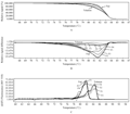
In this experiment, HRM analysis showed that SSR marker CH03d11 between ‘Fuji’ and ‘Telamon’ can arise similar DNA polymorphism melting curve in three kinds of different total reaction volume (Figure 1). This polymorphism changes can be shown by the view of three different forms: (1) the normalized and shifted melting curve (Figures 1A), which is adjusted all the samples in the context of the same temperature to compare the relative fluorescence signal values; (2) the normalized and temperature-shifted difference plot (Figures 1B), which selects one sample as the melting curves plotted baseline to highlight the relative differences in samples; (3) the melting peaks (Figures 1C), a derivative type, which is to compare the change rate of fluorescence signal between the samples with temperature, namely the fluorescence value and temperature. In addition, the peak corresponding to the specific temperature is the melting temperature (Tm) of the samples. The differences between samples can be obviously seen from the curve of the first graph, however, the second graph can definitely distinguish the differences in the samples, and we can easier to identify the samples. From the third graph, we can intuitively understand the difference in different samples’ melting temperature.
|
|
Seeing from Figure 1, the three different view forms can be clearly reflected the polymorphisms between the two cultivars, and the reproducibility of the experimental results is good in the different total reaction volume. As shown in Figure 1C, the change of reaction volume caused some deviations of the amplicon’s Tm value, fortunately, this bias did not affect the overall analysis effects.
1.2 Effect evaluation of different concentration of DNA for HRM analysis
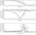
The concentration of DNA template is one of the main factors to affect PCR amplification. Generally, PCR amplification can normally carry out as DNA template concentration in a large range. It will result in no amplification product or produce non-specific amplification, while the template concentration is too high. When the template concentration is too low, it is also not conducive for effective PCR amplification. All these will directly affect the subsequent HRM signal detection. In this study, we set a gradient experiment for the DNA template concentration with 0.25 ng/μL, 0.5 ng/μL, 1.0 ng/μL, and 2.0 ng/μL in the total reaction of 5 μL. The result showed that the analysis of the samples is similar in the different DNA template concentration (Figures 2). That is to say, when the lowest concentration of DNA template is one-eighth of the highest, HRM can detect the PCR amplicon normally.
|
|
1.3 Effect evaluation of touchdown PCR protocol for HRM analysis
In the HRM reaction, it is very critical to select an appropriate annealing temperature (Mackay et al., 2008; Erali and Wittwer, 2010). HRM analysis effects were compared between the touchdown PCR protocol and the conventional PCR protocol. The result showed that these two kinds of amplified modes can get the same test results when the reaction volume was 20 μL, 10 μL and 5 μL, repectively (Figures 3).
|
|
2 Discussion
Currently, HRM technique has been widely adopted in human disease-related gene mutation scanning and molecular diagnostics fields, and formed a relatively complete research system (Luo et al., 2011). Recently, there are a few reports on HRM in plants. Such as, the identification of the different varieties of grapes and olives (Mackay et al., 2008); SNP marker analyses of expressed sequence tags from apple (Chagné et al., 2008); the identification of barley gene locus (Hofinger et al., 2009) and so on. Recently, Li et al (2011) have explored that the reaction system of HRM using rice as the test materials and the results showed that the stability of the volume of 5 μL reaction was worse than that of 10 μL, 20 μL reaction systems and caused some deviations of the amplicon’s Tm value. We also confirmed this phenomenon in apple. Previous reports that the volume system of HRM analysis is basically over 10 μL. But the scientists found that the volume reaction system of 5 μL was still good to polymorphism detection and genotyping, and other reagent consumption was one-fouth of 20 μL system recommended by the kit. The cost of HRM analysis has been reported as $1.50 per sample in 20 μL reaction system (Wu et al., 2008). Hence, without affecting the test results, 5 μL reaction system can greatly reduce costs. In addition, we also found when the DNA template concentration was 0.25 ng/μL, the HRM detection of the PCR amplicon can be carried out normally. Therefore, when the amount of DNA template is a little, we can consider a lower concentration in order to make sure the multiple experiments.
Touchdown PCR is a common analysis model in PCR amplification, its annealing temperature gradually dropped from a high value to a lower value with the amplification cycle. Then the following expansion cycle will be completed with this low value. This model can be normally amplified and reduce non-specific amplification.
The study showed that the touchdown PCR could get the same typing with the conventional PCR in the different reaction volume. Because touchdown PCR provided correct annealing temperatures for many primers at the same time, which would make it possible to amplify and analyze the multiple markers in the same run. It is important to improve the efficient use of the materials and the equipments.
3 Materials and Methods
3.1 Plant materials
Apple cultivars ‘Fuji’ and ‘Telamon’ were used in the study.
3.2 Genomic DNA extraction
DNA was extracted from fresh young buds (about 0.1 g) using modified CTAB method (Tian et al., 2003). DNA was quantified on a Ultrospec 3300 pro (Amersham Biosciences) and conserved at -20℃ until we needed.
3.3 PCR amplification and HRM verification
We used the SSR markers CH03d11 linkaged to apple columnar trait as the tested object. PCR amplification and HRM analysis were performed on the LightCycler® 480 II Real-Time PCR System in 96-well multi-well plates, and PCR reactions consisted of 1×LightCycler® 480 High Resolution Melting Master; supplemented with 2.0 mmol/L MgCl2 and 0.2 μmol/L each primer. We totally arrange three kinds of treatments for the reaction volume: 5 μL, 10 μL and 20 μL and four kinds of treatments for DNA template: 2 ng/μL, 1 ng/μL, 0.5 ng/μL and 0.25 ng/μL. The cycling program consisted of a universal PCR protocol as follows: pre-denaturalization for 10 minutes at 95℃, 95℃denaturalization for 10 seconds, 60℃ annealing for 15 seconds and 72℃ extension for 10 seconds. The amplification cycles were immediately followed by the high resolution melting steps of 95℃ for 1 minute, cooling to 40℃ for 1 minute, raising the temperature to 65℃and then raising the temperature to 95℃ with 25 fluorescent acquisitions per degree Celsius in this step. In addition, a touchdown protocol has a similar amplification cycles but with annealing temperatures decreasing from 60℃to 55℃ for 15 seconds. The annealing temperature decreased in subsequent cycles by 0.5℃ per cycle after the first 60℃ annealing step to 55℃ (Yin et al., 2011).
3.4 High resolution melting analysis
The melting curve was analyzed with the gene scanning software module (1.5 version) on the LightCycler® 480 II instrument.
Authors' contributions
CHW and MDB conceived the experimental design and objectives of all the HRM experiments, conducted the HRM data analyses, and wrote the manuscript. HY and JFL conducted a few data analyses and took an active part in experimental design method. CHW and YKT were the prime principal of the project and took part in reviewing and writing the manuscript. All authors have read and approved the manuscript.
Acknowledgements
This work was co-supported by Shandong province thoroughbred Project and Qingdao Municipal Science and Technology Program of Basic Research Projects (11-2-4-5-(4)-jch).
References
Chagné D., Gasic K., Crowhurst R.N., Han Yue-peng, Bassett H.C., Bowatte D.R., Lawrence T.J., Rikkerink E.H.A., Gardiner S.E., and Korban S.S., 2008, Development of a set of SNP markers present in expressed genes of apple, Genomics, 92(5): 353-358
http://dx.doi.org/10.1016/j.ygeno.2008.07.008 PMid:18721872
Chen B., Lei X.X., and Zhou X.M., 2009, Application of high resolution melting analysis in molecular diagnosis, Journal of Molecular Diagnosis and Therapy, 1(2): 120-124
Dong C., Vincent K., and Sharp P., 2009, Simultaneous mutation detection of three homoeologous genes in wheat by high resolution melting analysis and mutation surveyo, BMC Plant Biology, 9: 143 doi:10.1186/1471-2229-9-143
http://dx.doi.org/10.1186/1471-2229-9-143 PMid:19958559 PMCid:2794869
Erali M., and Wittwer C.T., 2010, High resolution melting analysis for gene scanning, Methods, 50(4): 250-261
http://dx.doi.org/10.1016/j.ymeth.2010.01.013 PMid:20085814 PMCid:2836412
Hofinger B.J., Jing H.C., Hammond-Kosack K.E., and Kanyuka K., 2009, High-resolution melting analysis of cDNA-derived PCR amplicons for rapid and cost-effective identification of novel alleles in barley, Theor. Appl. Genet., 119(5): 851-865
http://dx.doi.org/10.1007/s00122-009-1094-2 PMid:19578831
Li D., Wang J.Q., Bu D.P., Liu K.L., Li S.S., Zhao S.G., and Yu P. 2009, Study of high resolution melting and application, Biotechnology Bulletin, 7: 48-51
Li J.H., Wang X.H., Dong R.X., Yang Y., Zhou J., Yu C.L., Cheng Y., Yan C.Q., and Chen J.P., 2011, Evaluation of high-resolution melting for gene mapping in rice, Plant Mol. Biol. Rep., 29(4): 979- 985
http://dx.doi.org/10.1007/s11105-011-0289-2
Luo W.H., Guo ., Wang H., Liu Y.Z., Zhang J.G., and Chen Z.Q., 2011, Application of high resolution melting analysis in plant breeding, Chinese Agricultural Science Bulletin, 27(03): 10-14
Mackay J. F., Wright C.D., and Bonfiglioli R.G., 2008, A new approach to varietal identification in plants by microsatellite high resolution melting analysis: application to the verification of grapevine and olive cultivar, Plant Methods, 4(1): 8
http://dx.doi.org/10.1186/1746-4811-4-8 PMid:18489740 PMCid:2396621
Mader E., Lukas B., and Novak J., 2008, A strategy to setup codominant microsatellite analysis for high-resolution-melting-curve-analysis (HRM), BMC Genet., 9: 69
http://dx.doi.org/10.1186/1471-2156-9-69 PMid:18980665 PMCid:2588637
Norambuena P.A., Copeland J.A., KÅ™enková P., Štambergová A., and Jr M.M., 2009, Diagnostic method validation high resolution melting (HRM) of small amplicons genotyping for the most common variants in the MTHFR gene, Clinical Biochemistry, 42(12): 1308-1386
http://dx.doi.org/10.1016/j.clinbiochem.2009.04.015 PMid:19427845
Tan C.X., Luo J.F., and Ren Z.R., 2009, High resolution melting-new method for molecular diagnosis, Life Science Research, 13(3): 268-271
Tian Y.K., Wang C.H., Zhang J.S., Dai H.Y., and Chu Q.G., 2003, A RAPD marker of apple columnar gene (Co), Acta bot Boreal-Occident sin, 23(2): 2176-2179
Vossen R.H., Aten E., Roos A., and Dunnen J.T., 2009, High resolution melting analysis (HRMA)—more than just sequence variant screening, Hum. Mutat., 30(6): 860-866
http://dx.doi.org/10.1002/humu.21019 PMid:19418555
White H.E., Hall V.J., and Cross N.C.P., 2007, Methylation-sensitive high-resolution melting-curve analysis of the SNRPN gene as a diagnostic screen for Prader-Willi and Angelman syndromes, Clin. Chem., 53(11): 1960-1962
http://dx.doi.org/10.1373/clinchem.2007.093351 PMid:17890436
Wittwer C.T., Reed G.H., Gundry C.N., Vandersteen J.G., and Pryor R.J., 2003, High-resolution genotyping by amplicon melting analysis using LCGreen, Clinical Chemistry, 49(6): 853-860
http://dx.doi.org/10.1373/49.6.853 PMid:12765979
Wojdacz T.K., and Dobrovic A., 2007, Methylation-sensitive high resolution melting (MS-HRM): a new approach for sensitive and high-throughput assessment of methylation, Nucleic Acids Research, 35(6) : e41- doi:10.1093/nar/gkm013
Wu S. B., Tavassolian I., Rabiei G., Hunt P., Wirthensohn M., Gibson J. P., Ford C. M., and Sedgley M. 2009, Mapping SNP-anchored genes using high-resolution melting analysis in almond, Mol. Genet Genomics, 282(3): 273-281
http://dx.doi.org/10.1007/s00438-009-0464-4 PMid:19526371
Wu S. B., Wirthensohn M. G., Hunt P., Gibson J. P., and Sedgley M., 2008, High resolution melting analysis of almond SNPs derived from ESTs, Theor. Appl. Genet., 118(1): 1-14
http://dx.doi.org/10.1007/s00122-008-0870-8 PMid:18781291
Zhou L., Vandersteen J., Wang L., Fuller T., Taylor M., Palais B., and Wittwer C.T., 2004, High-resolution DNA melting curve analysis to establish HLA genotypic identity, Tissue Antigens, 64(2): 156-164
http://dx.doi.org/10.1111/j.1399-0039.2004.00248.x PMid:15245370
. PDF(390KB)
. FPDF(win)
. HTML
. Online fPDF
Associated material
. Readers' comments
Other articles by authors
. Mudan Bai
. Caihong Wang
. Hao Yin
. Yike Tian
. Jiefa Li
Related articles
. Apple ( Malus domestica )
. HRM
. Reaction volume
. DNA template concentration
. Touchdown PCR
Tools
. Email to a friend
. Post a comment


