Research Article
Assessment of Microbiological and Keeping Quality of Ocimum Gratissimum and Vernonia Amygdalina Crude Extract on Sarotherodon Galilaeus Snacks 
2 Department of Fisheries and Aquaculture, Adekunle Ajasin University, Akungba Akoko, Ondo State, Nigeria
3 Department of Microbiology, Adekunle Ajasin University, Akungba Akoko, Ondo State, Nigeria
 Author
  Correspondence author
Bioscience Methods, 2017, Vol. 8, No. 2 doi: 10.5376/bm.2017.08.0002
Received: 15 Sep., 2017 Accepted: 26 Oct., 2017 Published: 21 Nov., 2017
Ileogben T.A., Oginni O., Olatunbosun A., Olajubu F.A., Omobuwa M.O., Ojo B.O., Umeh E.S., and Ayodeji O.O., 2017, Assessment of microbiological and keeping quality of Ocimum Gratissimum and Vernonia Amygdalina crude extract on Sarotherodon Galilaeus snacks, Bioscience Methods, 8(2):18-30 (doi: 10.5376/bm.2017.08.0002)
Evaluation of antimicrobial and keeping qualities potentials of Ocimum gratissimum (OGE), Vernonia amygdalina (VAE), and Ocimum gratissimum plus Vernonia amygdalina (OVE) crude extracts on fish snack made from Sarotherodon galilaeus, was carried out to determine their for suitability for food preservation. Twelve fish samples grouped into three class sizes (A, B, C), were used. They were gutted, filleted, cut into pieces, staked on palm frond sticks, and fortified with extracts, spiced and dried with a smoldering fire. The snacks were allowed to cool and kept in Aluminum foil, Brown paper and polythene sheet. Snacks analysis for microbial and keeping qualities was carried out using NA agar for bacteria and PDA for fungi using standard methods. Data collected were subjected to ANOVA using SAS® 2008 version 9.1. Mean pH value ranged from 9.60±0.1 to 11.10±0.15 for freshly produced snack, untreated snacks had the least mean pH value (9.60±0.1) and the highest mean bacteria count (9x10²±2.65). Fungi counts (5x10²±2.08 to 8x10²±6.08) were not significantly different between the samples. It was observed that snacks treated with Ocimum gratissimum crude extract had the highest pH value and the lowest bacteria and fungi load; hence it was more effective for fresh snack preservation. Five bacteria and eight fungi were isolated and identified. Result of pH and microbial analysis after storage showed that the pH values ranged from 9.50±0.00 to 10.17±0.06. For treated snacks, snacks packaged in aluminum foil had the least pH value and the highest microbial load, snacks packaged in brown paper had the highest pH value and the highest microbial load. For packaged snacks, snacks treated with the mixture of Ocimum gratissimum and Vernonia amygdalina cude extraxts was more effective as it had the lowest bacteria and fungi load. Microbiological analysis revealed that Staphylococcus aureus and Aspergillus spp were the dominant bacteria and fungi isolated; however, their counts were within the acceptable limit of International Commission on Microbiological Specification. Polyethylene bag gave the best result for packaging materials as it showed no deleterious quality change in the snacks. Addition of plants extract and composite spice to the fish fillet had effect on the microbial and shelf life of the product. Key Words; Fish snack, Microbiological analysis, Ocimum gratissimum, Sarotherodon galilaeus, Vernonia amygdalina.
Background
Despite the high degree of awareness of food preservation methods, there is increasing occurrence of food loss/disease outbreaks caused by spoilage and pathogenic microorganisms in foods. Due to consumer awareness and negative perception of artificial preservatives in food, in recent years attention is shifting toward alternatives that the consumers recognize as natural. In recent years, consumers prefer fewer chemicals and more natural foods, thus, there is growing interest in using natural antimicrobial compounds, including extracts of herbs and spices, as salt replacers or alternatives to synthetic compounds for food preservation (Witkowska, 2013). Plant extracts are now getting more space in food industry to prevent the propagation of bacteria that affect the spoilage of food or the spread of so-called food-borne diseases. The conditions of storage can also cause physiological spoilage. Various methods exist to ensure that food maintain their quality long after harvesting (Oladotun and Deborah, 2013). The major shortcoming posed by the use of many antimicrobials is the chemical residues it deposits in the food. The use of plants in preservation has been in use for a long period, though little understood then, it served its purposes. A better understanding of the roles of plant parts in increasing the shelf life of farm produce has helped in the creation of more effective and safer means of pest and microbial control (Oladotun and Deborah, 2013).
Ocimum gratissimum (L.) commonly known as scent leaf is an herbaceous, perennial shrub of the family Lamiaceae, often found growing around villages in Nigeria with its characteristic smell for seasoning dishes (Okujagu, 2008). Owalade et al. (2000) reported that leaf extracts of O. gratissimum and Verononia amygdalina effectively protected maize seeds from seed borne infection of Fusarium moniliforme. Also Okoi and Afuo (2009) reported that crude extracts of O. gratissimum effectively exhibited antifungal activity on Cercospora arachidicola, the causal organism of leaf spot disease of groundnut.
Vernonia amygdalina, variously known as bitter leaf is a tropical shrub, which range from 1-3 m in height with petiole leaf of about 6mm in diameter, and elliptic in shape (Igile et al., 1995). The leaves are dark green coloured with a characteristic odour and a bitter taste. The species is indigenous to tropical Africa and is found wild or cultivated all over sub- Saharan Africa (Bosch et al., 2005). All parts of the plant are pharmacologically useful. Both the roots and leaves are used in phyto-medicine to treat fever, hiccups, kidney disease and stomach discomfort, among others (Gill, 1992; Hamoiona and Saffaf, 1994). Anti-helmitic and anti-malarial properties (Abosi and Raserika, 2003) as well as anti-tumourigenic properties (Izevbigie et al., 2004) have also been reported for extracts from the plant. Other studies have demonstrated hypoglycaemic and hypolipidaemic effects of the leaf extract in experimental animals (Akah and Okafor, 1992; Nwanjo, 2005). The study is therefore aimed at providing information on the anti-microbial potentials of Ocimum gratissimum and Vernonia amygdalina crude extract on Sarotherodon galilaeus snacks.
1 Materials and Methods
1.1 Source of raw materials
The samples used for this study were taken from sample used for nutritional assessment of fish snack made from Sarotherodon galilaeus. 940 grams of Vernonia amygdalina and 827 gms of Ocimum gratissimum leaves and every other ingredient for composite spice “suya spices” and other materials used were purchased from Akungba Akoko, Ondo state, Nigeria.
1.2 Production of spices
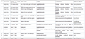
The spice was prepared from the following ingredients; Human edible Groundnut cake locally called “kulikuli”, Ginger, Garlic, Cayenne pepper flakes, Black Pepper, Cooking salt and African Negro pepper. These ingredients were ground into powdery form to increase surface area and improve attachment. The spice was similar to “Yaji” that is “suya” spice (Table 1).
|
Table 1 Spice composition |
.png)
1.3 Preparation of crude extracts and brine plus crude extract
Nine hundred and forty grams of Vernonia amygdalina and 827 gms of Ocimum gratissimum leaves were quickly washed in jet of tap water and rid of excess water before squeezing into separate bowls to obtain crude extract. The extracts were diluted to acceptable taste based on taste range finding test. Ocimum gratissimum was diluted with potable water to give 54.5% v/v concentration; also, Vernonia amygdalina was diluted with potable water to give 23 v/v concentration while equal volume of Vernonia amygdalina and Ocimum gratissimum extract mixed together was diluted to give 45.5 v/v concentration. 8 grams of salt was added to Vernonia amygdalina extract, 6 gms of salt to Ocimum gratissimum extract and eight gms of salt into mixture of Vernonia amygdalina and Ocimum gratissimum extract for brining. A five man taste panelists was used to determine the acceptable level of extract and salt concentration.
1.4 Preparation of fish snack
The fish samples were washed in jet of tap water to remove dirt and thereafter, allowed to drain. The fish were scaled, gutted and washed clean again after gutting to prevent contamination of the muscle. Thereafter, they were filleted, and the weight of the fillets taken. The fillets were cut into smaller pieces (2.5 cm x 6 cm), and the pieces were staked on sterilized palm frond sticks. Staked fillets pieces were weighed and soaked in brine and mixture of plant extracts and brine for two minutes, the brined/ brine plus extracts stakes were hung to drain excess solution after which oil and spice was applied. Smouldering fire from wood and charcoal combustion was used to cook dry the snacks.
1.5 Keeping quality assessment
The snacks were packaged in three different sterilized packaging materials (Polythene bag, Aluminum Foil and Brown Paper). Snacks were kept under ambient condition after packaging to determine the shelf life and appropriate packaging material for a period of ten (10) days based on the shelf life of some commercial snacks such as sausage roll e.g. Gala, Beefy etc. Each of the packaged snacks was wrapped with brown paper and the untreated sample was kept in a brown paper without either of the other packaging material. A representative sample of the snack was checked/examined daily for microbial or sensory loss during the period of storage.
1.6 pH determination
One gram of each sample was ground and poured into 9 mls of distilled water. The mixture was homogenized by gently shaking the container of mixture (sample and distilled water). SURGIFIELD® hand held pH meter (Model; SM-602A) was dipped into the mixture for 3 minutes and the pH value determined in triplicate.
1.7 Microbial analysis
1.7.1 Sterilization of materials
The test tube bottles, conical flasks and all glassware used for this work were thoroughly washed with household detergent, rinsed with clean water, air dried and sterilized in the hot air oven at 170°C for 2 hours.
1.7.2 Media preparation
Nutrient Agar and Potato Dextrose Agar were prepared according to the manufacturer`s (Oxoid®) specification by weighing (2.8 gms of Nutrient Agar and 3.9 gms of Potato Dextrose Agar into 100 mls of water). 8.4 gms of Nutrient Agar and 11.7 gms of Potato Dextrose Agar were weighed and poured into two conical flasks, 300 mls of distilled water was poured into each of the flasks and mixed by shaking the flasks with the opening corked with cotton wool and sealed with foil paper, after shaking the flasks, the flask containing the mixture were autoclaved at 121°C for 15 mins after which the autoclave was allowed to cool down, the flasks containing the media mixtures were then removed and allowed to cool to about 45°C before pouring into plate.
1.7.3 Innoculation of samples
One gram each of the snack sample in powdered form was serially diluted in three test tubes containing 9 mls of sterile distilled water to give dilution 10-2 and 10-3 respectively. Hands and work table were sterilized with ethanol; 0.5 mls of the 10-2 dilution sample was poured into the petridishes in triplicate and labeled accordingly. After pouring, the petridishes were manually rocked gently both clockwise and anti-clockwise for the media to form a uniform layer. The petridishes were allowed to cool for one hour for the media to solidify, taped and kept in the incubator at a temperature of 37°C and observed after 24 hours.
For fungi, the Potato Dextrose Agar was also poured into the petridishes containing the 10-2 dilution of the snacks sample in triplicate, the plates were gently rocked and allowed to cool and solidify before taping the plates. The plates were observed for 7 days for fungi growth.
1.7.4 Enumeration of microbial colonies
Colony counting was carried out visually by counting the number of visible colonies that appeared on the plates. Calculation of colony forming unit (CFU) per ml for the bacteria and the spore forming unit (SFU) per ml for the fungi was based on the formula:
.png)
1.7.5 Identification of bacteria and fungi
The identification of bacteria and fungi was based on the cultural characteristics (shape, colour of the pigment, opacity, elevation, edge of colony, consistency and surface), staining reaction (gram staining and lacto phenol cotton blue staining), and the biochemical tests (nitrate reduction, indole production, urease, coagulation and sugar fermentation) as reported by Cowan and Steel, (2002) (Famurewa et al., 2009).
1.7.6 Cultural characteristics of the bacteria
The bacterial isolates were streaked for single colonies, incubated at 37°C for 24 hours and observed directly on Agar plates to note the shape, size, colour, of pigment, opacity, elevation surface and edge of the colony.
1.7.7 Gram staining for bacteria
The gram staining steps include, the preparation of a heat fixed smear from an 18-24 hour culture, which was stained with crystal violet for one minute before the solution was poured off and rinsed off with gram iodine solution, the iodine was allowed to react for one minute then poured out and the slides washed with 95% alcohol until no more violet comes out from the slides. The slides were rinsed under a gentle running tap water and counter stained with safranin for 1 minute, the slides were blotted dry and examined with microscope (LXSZ-107BN: England) under oil immersion lens to observe the morphology of the cells.
1.7.8 Fungi staining
Few drops of lactophenol cotton blue solution were placed on slides, the inoculating needle was flamed and swirled for 20 seconds, after which, it was used to transfer fungus onto the slides, emulsified and the organisms covered with a cover slip. The prepared slides were viewed under microscope (LXSZ-107BN: England) and the fungi were observed.
1.7.9 Spore staining
The ability of bacteria isolates to produce endospores was determined. A heat smear was prepared from an 18-24 hours culture; malachite green solution was added and steamed for 5-10 minutes. The slides were washed with water, blotted dry and examined under the microscope (LXSZ-107BN: England) with an oil immersion objective lens; spores were stained green while bacteria cells were stained red.
1.8 Biochemical test
1.8.1 Catalase test
The tested organisms were inoculated onto Nutrient Agar plates and incubated at 37°C for 24 hours. 3% volume concentration of hydrogen peroxide was placed on a colony, emulsified and examined for oxygen bubbles which indicate the presence of catalase.
1.8.2 Coagulase test
An 18-24 hours culture was used for this test. A loop full of normal saline was placed on each section of a slide and was emulsified with a small amount of the 18-24 hours old culture until a homogenous suspension was obtained.
A drop of human plasma was added to one of the suspensions and stirred for 5 seconds with the other suspension as a control. A coagulase positive result was indicated by clumping which did not re-emulsify.
1.8.3 Motility test
Motility media was prepared according to the manufacturer`s (Oxoid ®) specification by weighing (53 g per Litre: England), this was used to demonstrate the motility of organism due to flagella. 18-24 hours old organism was stabbed on the agar and incubated at 37°C for 24-48 hours. Motile bacteria were identified by their movement through the stabbed agar from top to bottom.
1.8.4 Urease test
A little of the culture of the test bacteria was streaked over the surface of the agar slant of urease test medium with phenol red as indicator and incubated at 37°C for 7 days. A control of the basal medium containing no added urea was equally inoculated. A colour change of the medium from yellow to pink or red indicated a positive result and no colour change indicate a negative result (Fawole and Oso, 2001).
1.8.5 Oxidase test
A piece of filter paper was placed in a sterile Petri dish and 2-3 drops of freshly prepared oxidase reagent was added. Using a sterile glass rod, a colony of the test bacterium was picked and smeared on the filter paper and observed for 10 seconds. The presence of blue-purple colour indicates a positive oxidase while absence of blue-purple colour indicates a negative oxidase test (Fawole and Oso, 1998).
1.8.6 Tests for indole production
A young culture of the test organism was inoculated into 10 mls peptone water in test tubes and incubated at 37°C for 7 days, Kovac`s reagent (0.5 mls) was added to each test tube and the test tubes were shaken gently and allowed to stand. A rose-purple colour developed in the presence of indole Famurewa et al. (2009).
1.8.7 Tests for sugar fermentation
A 1.0% NaCl, fermentable sugar and 0.01% phenol red were used to prepare the medium. The medium (5 mls) was dispensed into test tubes containing inverted Durham`s tubes after which they were sterilized at 115°C for 15 minutes. The test tubes were inoculated with 18-24 hour old culture of the test organisms and incubated at 37°C for 5 days. The test tubes were observed and the presence of yellow colour instead of red indicates fermentation Famurewa et al. (2009).
1.8.8 Statistical analysis and data presentation
Data were subjected to one-way analysis of variance (ANOVA) and the means separated using Duncan`s Multiple Range Test. Statistical Analysis System (SAS®), version 9.1 package was employed for statistical analysis at 5% confidence level. Data were presented in mean±, percentile, as well as frequency, pie and bar chats as appropriate.
2 Results
2.1 Microbial qualities
The result for pH and microbial analysis of freshly produced snack is presented in Table 2. It showed that the mean pH value ranged from 9.60±0.1 – 11.10±0.15; mean bacteria count ranged from 1x10²±1.15 – 9x10²±2.65 cfu/g and fungi mean count ranged from 5x10²±2.08 – 8x10²±2.89 sfu/g.
|
Table 2 pH and microbial counts of freshly produced snack before packaging for storage Note: Means with the same superscript are not significantly different (p>0.05) |
.png)
The untreated snacks had the least mean pH value (9.60±0.1) and the highest mean bacteria count 9x10²±2.65 cfu/g which were significantly different.
Snacks treated with Mixture of Vernonia amygdalina plus Ocimum gratissimum crude extract had significantly highest pH value 11.10±0.15 its bacterial count was the same with snacks treated with Ocimum gratissimum crude extract 1x10²±1.15 cfu/g as the least.
For fungi, mean count for untreated snacks and snacks treated with Vernonia amygdalina plus Ocimum gratissimum crude extract were the same (8x10²± 2.8 sfu/g). Fungi counts were not significantly different between the samples.
Five bacteria and eight fungi species were isolated and identified as shown in Table 3 and Table 4.
|
Table 3 Morphological, physiological and biochemical characteristics of bacteria isolated from freshly produced snack before packaging Note: + = Positive; - = Negative |
.png)
|
Table 4 Morphological characteristics and suspected fungi organism isolated from freshly produced snack before packaging |
.png)
2.2 Keeping quality
Keeping quality of the snacks in three packaging materials varied with treatments. Physical observation at the end of keeping/ storage time showed little changes in colour and presence of mould growth as shown in Table 5.
|
Table 5 Physical observation of packaged snacks after ten (10) days of storage |

Result of pH and microbial analysis after storage presented in Table 6 showed that the pH values ranged from 9.50±0.00 to 10.17±0.06, Bacteria mean count ranged from 4x10² cfu/g±2.00 to 14x102±3.00 and Fungi from 7x10² sfu/g±1.53 to 16x102±3.51.
|
Table 6 pH and microbial counts of packaged snack after ten days (10) of storage Note: Means with the same superscripts are not significantly different (p>0.05) |
.png)
Analysis of the results for snack treated with crude extracts showed that the mean pH, bacteria and fungi counts for those treated with mixture of Vernonia amygdalina plus Ocimum gratissimum ranged from 9.6±0.15 to 10.07±0.12, 7x10² cfu/g±1.00 to 10x102 cfu/g±1.53 and 7x10² sfu/g±1.53 to 10x102 sfu/g±0.58 respectively. For snacks treated with Vernonia amygdalina crude extract, the mean pH ranged from 9.57±0.06 to 9.93±0.06, bacteria mean count from 5x10² cfu/g±1.53 to 13x102 cfu/g±5.51 and fungi mean count from 9x10² sfu/g±1.53 to 11x102 sfu/g±4.00. The mean pH, bacteria and fungi counts for Ocimum gratissimum treated snacks ranged from 9.50±0.00 to 9.53±0.15, 11x102 cfu/g±1.53 to 14x102 cfu/g±3.00 and 7x10²±2.00 sfu/g to 16x102±3.51 sfu/g respectively. The untreated snack had mean pH, bacteria and fungi counts of 9.97±0.12 to 10.17±0.06, 4x10² cfu/g±2.00 to 12x102 cfu/g±5.00 and 9x10² sfu/g±2.52 to 13x102 sfu/g±1.00 respectively.
For keeping quality, the result for the different packaging materials showed that snack packaged in aluminum foil had the least pH value 9.6±0.15 and recorded the highest bacteria and fungi count; 10x102 cfu/g±1.53 and 10x102 sfu/g±0.58 respectively for snack treated with mixture of Vernonia amygdalina plus Ocimum gratissimum crude extract. For snack treated with Vernonia amygdalina crude extract, the highest pH value was recorded for snacks kept in polythene bag 9.93±0.06 with significant difference and also had the least count for both bacteria and fungi 5x10² cfu/g±1.53 and 9x10² sfu/g±1.53 respectively.
The least pH was recorded for aluminum foil packaging ranging from (9.50±0.00 to 9.6±0.15) although with no significant difference and had (p>0.05) bacteria count ranging from (10x102±1.53 to 14x102 cfu/g±3.00) and fungi count ranging from (10x102 to 16x102 sfu/g±3.51) with significant difference for all the treatments.
Brown paper packaging recorded the highest pH value 10.17±0.06 with significant difference. It recorded the highest bacteria count 12x102 cfu/g±5.00 while Polythene bag had the lowest pH value 9.97±0.12 and the highest fungi mean count 15x102 sfu/g±3.00 with significant difference for snacks without fortification.
Six bacteria and thirteen fungi were isolated and identified at the end of the storage period (Table 7).
|
Table 7 Morphological characteristics and suspected fungi organism isolated on packaged snack after storage |
The occurrence of microbes in the snack showed that both transient and resident strains were present. Occurrence of microbes in the packaged snacks is presented in Figure 1 and Figure 2. The occurrence for bacteria revealed that Staphylococcus aureus was the dominant organism, accounting for 53% of the total bacterial load. Seven percent occurrence recorded for Bacillus subtilis was the least for bacteria. The untreated snack had the highest microbial diversity. Of importance is the presence of Pseudomonas spp which is a common spoilage bacterium in the sample. The dominant fungus in the sample is Aspergillus spp and it accounted for 45% of fugal population in the samples. For fungi, the two percent occurrence recorded for Trichoderma longibrachiatum, Candida sake, and yeast was the least.
|
Figure 1 Percentage occurrence of bacteria isolated from snack samples |
.png)
|
Figure 2 Percentage occurrence of fungi isolated from snack samples |
.png)
3 Discussion
Alkalinity/ acidity of foods affects quality and shelf life because different microbes have different optimum pH, the pH of snacks treated with the crude extracts and the untreated was analyzed for the effects on microbial presence and diversity (Table 8). The untreated snack had the least pH (9.60±0.1) and highest counts for bacteria and fungi loads, Vernonia amygdalina and Ocimum gratissimum crude extracts had effect on the microbial load but Ocimum gratissimum had more effect on both bacteria and fungi load. The mixture of Vernonia amygdalina plus Ocimum gratissimum reduced bacterial load but had no effect on fungi growth in the freshly produced snack, from the result of this study, it could be deduced that there may be an antagonizing effect of some phytochemicals in the plants on freshly produced snacks. Ocimum gratissimum had the best effect in reducing bacteria and fungi loads; and could be a suitable substitute to synthetic preservatives for the preservation of the fish snacks and probably other processed fish products. Three genera of bacteria, Bacillus spp, Micrococcus spp and Staphylococcus were isolated from both freshly produced and packaged snack. Micrococcus species are strictly aerobic Gram positive cocci bacteria that can contaminate food product through water, dust and handler. Staphylococcus aureus as a Gram positive bacterium is a facultative anaerobe which can grow in both aerobic and anaerobic conditions this probably might be responsible for its being the dominant bacterium, its presence in the snack could be from atmosphere, water and handlers. The counts of the isolated organisms were still lower than the limit of 103 cfu/g recommended by ICMSF (2002) in Good Manufacturing Practice (GMP). This could be attributed to the high hygienic standard observed during the processing, and an assurance of the safety of freshly produced S. galilaeus snack consumption.
|
Table 8 Microorganisms isolated from different snack samples Note: A =Untreated; B = Ocimum gratissimum fortified snack; C = Vernonia amygdalina fortified snack; D = Mixture of Vernonia amygdalina plus Ocimum gratissimum fortified snack |

Eight species of fungi, Mucor plumbeus, Rhizopus stolonifer, Alternaria tenuissima, Aspergillus niger, Fusarium solani, Penicillium chrisogenum, Trichoderma longibrachiatum and Yeast Candida sake were isolated from freshly produced snack while thirteen species of fungi, Mucor plumbeus, Rhizopus stolonifer, Alternaria tenuissima, Aspergillus niger, Fusarium solani, Penicillium chrisogenum, Trichoderma longibrachiatum, Yeast, Aspergillus fumigatus, Fusarium verticilliodes, Mucor hiemalis, and Aspergillus flavus, were isolated from packaged snacks. The trend in microbial load of packaged snacks indicated that the lower the pH values the higher the microbial counts. Snacks packaged in aluminum foil had the highest microbial load and least pH values in all the treatments while Polythene had the least microbial load and fluctuating pH values, this is similar to Ogunsola and Omojola (2008) observation for beef and pork kilishi. The higher microbial load in aluminum foil might be, due to absence of pore in the material, and resultant buildup of moist, warm, a favourable environment for the spore producing microbes to sporulate under ambient condition. Staphylococcus aureus is normal skin flora of 25% healthy human and it does not cause diseases unless mixed with foods. Staphylococcus aureus is significant among staphylococcus genus as it can produce different types of toxins which are heat resistant. Staphlococcus aureus was the most prominent spoilage organism isolated; they survived in all the samples analyzed. Pearson (1986) reported Staphlococcus aureus as a Gram positive bacterium, its toxin causes food poisoning and it is resistant to heat, radiation and drying. Evans et al. (1983) reported S. aureus as a major cause of food poisoning and as a facultative anaerobe can grow under both aerobic and anaerobic conditions. Although it was dominant in all samples, however, its level was still within the range of satisfactory microbial load for human health (ICMSF, 2002). Pseudomonas spp are known to be spoilage bacteria, especially P. aeruginosa and P. fluorescens have been considered as opportunistic pathogenic species (Altinok et al., 2006). The presence of P. aeruginosa could be attributed to its being part of normal floral of fresh water fishes and its inherent spores which sporulate in favourable condition.
Mould thrives better in medium with high moisture content (Pearson, 1986). The presence of Aspergillus spp and Mucor spp which are spoilage fungi could be as a result of moist environment created by Aluminum foil packaging material which favoured their growth and consequent high population in the sample.
Physical observation of snacks after the period of ten days storage in the different packaging materials revealed that Polythene was the best packaging material for the snacks as no change was observed in snacks packaged in polythene. Snacks packaged in aluminum foil had varying degrees of colour change due to presence of mould, this could be due to absence of pore in aluminum foil which might have allowed heat and moisture to buildup thus creating favourable environment for mould growth.
In this study, polythene was the best packaging material in terms of physical and microbial qualities, snacks were microbiologically safe for consumption within ten days of production the microbial load were below the limit (103 cfu/g) recommended by ICMSF in Good Manufacturing Practice (GMP).
Acknowledgements
My gratitude goes to my Maker, best friend, Comforter, hope, saviour, guider, Jesus Christ for the grace bestowed on me to complete this work. I give gratitude to Dr. O. Oginni for his love, selfless contributions to the success of this work. I appreciate Akele Olatunbosun, for his effort and guidance during the microbial bench-work. I also appreciate Dr. Festus A. Olajubu for reading and effecting necessary corrections. I will not fail to appreciate my loving and supportive parents Mr. and Mrs. Ileogben for their prayers, provision, care and support throughout the research work.
Authors’ contributions
Tina Agbon Ileogben is the main owner of the thesis work. Festus. A. Olajubu is a co-supervisor from the cognate department of micriobiogy and contributed by reading and making sure that proper micriobiology terms and rules are used and followed. Akele Olatunbosun is the technologist who worked with me and assisted throughout the practical period. Olatunde Oginni is the supervisor of Tina Agbon Ileogben, supervised, read and made corrections throughout the research period. Omobuwa Mary Oluwaseun, Ojo Blessing Olusoji, Umeh Eberechukwu Stella, and Ogunojemite Oladotun Ayodeji are undergraduate students who assisted during the practical period.
Abosi A.O., and Raseroka B.H., 2003, In vivo antimalarial activity of Vernonia amygdalina, Br. J. Biomedical Science, 60: 89-91
https://doi.org/10.1080/09674845.2003.11783680
Akah P.A., and Okafor C.I., 1992, Hypoglycemic effect of Vernonia amygdalina (Del) in experimental rabbits, Plant Med. Res, 1: 6-10
Altinok I.S., Kayis and Capkin E., 2006, Pseudomonas putida infection in rainbow trout, Aquaculture, 261(3): 850-855
https://doi.org/10.1016/j.aquaculture.2006.09.009
Bosch C.H., Borus D.J., and Siemonsma J.S., 2005, Vegetables of Tropical Africa, Conclusions and Recommendations Based on PROTA 2: ‘Vegetables’, PROTA Foundation, Wageningen, Netherlands, 10 modules, pp.68
Evans J.B., Ananaba G.A., Prate C.A., and Bergdoll M.S., 1983, Enterotoxin Production by a typical Staphylococcus aureus from poulty, Journal of Applied Bacteriology, (54):527
Famurewa O., and David O.M., 2009, Cell Phone: A Medium of Transmission of Bacterial Pathogens, Marsland Press World Rural Observations, 1(2): 69-72
Fawole M.O., and Oso B.A., 1998, Laboratory Manual of Microbiology, Spectrum Books Limited, Ibadan, pp.126
Fawole M.O., and Oso B.A., 2001, Laboratory Manual of Microbiology, Spectrum Books Limited, Ibadan, pp.127
Gill L.S., 1992, Ethnomedical Uses of Plants in Nigeria. Uniben Press, Benin City, Nigeria, pp.243
Hamowia A.M., and Saffaf A.M., 1994, Pharmacological studies on Vernonia amygdalina (Del) and Tithonia diversifolia (Gray), Vet. Med. Giza, 2: 91-97
ICMSF, 2002, International Commission on Microbiological Specifications for Foods, Microorganisms in foods, Microbiological testing in Food Safety Management, Kluwer Academic/Plenum Publishers, New York, pp.199
Igile G.O., Oleszyek W., Burda S., and Jurzysta N., 1995, Nutritional assessment of Vernonia amygdalina leaves in growing mice, J. Agric. Food Chemistry, 43: 2126-2166
https://doi.org/10.1021/jf00056a038
Izevbigie E.B., Bryant J.L., and Walker A., 2004, A novel natural inhibitor of extracellular signal related kinases and human breast cancer cell growth, Experimental Biol. Med (Maywood), 229: 163-169
https://doi.org/10.1177/153537020422900205
Nwanjo H.U., 2005, Efficacy of aqueous leaf extract of Vernonia amygdalina on the plasma lipoprotein and oxidative status of diabetic rat models, Nigerian Journal of Physiological Sciences, 20: 39-42.
Ogunsola O.O., and Omojola A.B., 2008, Qualitative evaluation of ‘Kilishi’ prepared from beef and Pork, African Journal of Biotechnology, 7 (11): 1753-1758
https://doi.org/10.5897/AJB08.354
Okujagu T.F., 2008, Medicinal plants of Nigeria, South East Zone Vol. 1 Nigerian National Medicinal Development Agency, Federal Ministry of Science and Technology Lagos, Nigeria, pp.180
Okoi A.I., and Afuo C.O., 2009, Effect of leaf extracts of three plant species on Cercospora arachidicola Hori, the causal fungus of leaf spot disease of groundnut (Arachis hypogea L), Nig., Journal of Food Research, pp.99
Oladotun A.F., and Deborah A.O., 2013, Use of Plant Antimicrobials for Food Preservation World Academy of Science, Engineering and Technology, International Journal of Biological, Biomolecular, Agricultural, Food and Biotechnological Engineering, 7(12):1
Owalade B.F., Amusa F.A.N., and Osikanlu Y.O.K., 2000, Efficacy of certain indigenous plant extract against seed-borne infection of Fusarium moniliforme on maize (Zea mays) in south western Nigeria, Cereal Research Communication, 28: 323-324
Pearson M.A., 1986, Advances in Meat Research; Meat and Poultry Microbiology, McMillan Publishers Limited, England, pp.195
https://doi.org/10.1007/978-1-349-09145-4
Witkowska A.M., Hickey D.K., Alonso-Gomez M., and Wilkinson M., 2013, Evaluation of antimicrobial activities of commercial herb and spice extracts against selected food-borne bacteria, Journal of Food Research, (2): 37-54. Jour. Plt. Protect 22. Special edition, pp.132-139


. PDF(452KB)
. FPDF(win)
. HTML
. Online fPDF
Associated material
. Readers' comments
Other articles by authors
. Tina Agbon Ileogben
. Olatunde Oginni
. Akele Olatunbosun
. Festus. A. Olajubu
. Mary Oluwaseun Omobuwa
. Blessing Olusoji Ojo
. Eberechukwu Stella Umeh
. Ogunojemite Oladotun Ayodeji
Related articles
. Fish snack
. Microbiological analysis
. Ocimum gratissimum
. Sarotherodon galilaeus
. Vernonia amygdalina λ
Tools
. Email to a friend
. Post a comment