Research Report
Tea Seed Oil Restores Blood Pressure, Redox Balance and Lipid Homeostasis in L-NAME-Induced Hypertensive Rats 
2 Department of Veterinary Physiology, Biochemistry and Pharmacology, Faculty of Veterinary Medicine, University of Ibadan, Nigeria
 Author
  Correspondence author
Bioscience Methods, 2026, Vol. 17, No. 3
Received: 17 May, 2026 Accepted: 18 Jun., 2026 Published: 29 Jun., 2026
Background: Hypertension remains a leading cause of cardiovascular disease and mortality worldwide. This study explores the potential of tea seed oil (TSO) in mitigating hypertension-related complications in a rat model induced with N(G)-nitro-L-arginine methyl ester (L-NAME). TSO was extracted following standard protocols, and experimental groups (A - E) were administered L-NAME along with varying doses of TSO or Enalapril Maleate (as a positive control) for 28 days. Blood pressure indices were measured non-invasively, and blood and tissue samples were collected for biochemical analyses.
Results: Groups B and E showed significant changes in systolic blood pressure (SBP) compared to the control (Group A). Additionally, SBP decreased notably in groups C, D, and E compared to hypertensive group B. The administration of TSO at the highest dose (0.6 ml/kg) caused a reduction in diastolic blood pressure (DBP) in group D, similar to the effect of enalapril maleate in group E. Group B, subjected only to L-NAME administration, exhibited notable increases in LDL-C. Liver function markers like AST, ALT, and ALP showed significant changes across groups, with TSO administration leading to reductions in AST in groups C and D, and in group E compared to group B. Group B rats showed a significant rise in malondialdehyde (MDA) compared to Group A. TSO administration in Groups C and D slightly reduced MDA towards Group A levels. In Group E, MDA significantly differed from both groups A and B. Catalase (CAT) decreased significantly in group B but remained unchanged in groups C, D, and E compared to A or B. Superoxide dismutase (SOD) decreased significantly in group B compared to A but increased in C, D, and E compared to B. Glutathione peroxidase (GPx) decreased in groups B and C compared to A but increased in C compared to B. groups D and E showed no significant difference in GPx values compared to either A or B. Reduced glutathione (GSH) increased significantly in TSO-treated Groups C and D and in the standard drug group compared to A or B.
Conclusions: These findings highlight the therapeutic potential of TSO as a natural adjunct in the management of hypertension and its related complications.
1 Background
Hypertension remains the leading risk factor for cardiovascular diseases, and its annihilative effect is felt in the form of reduced life expectancy and premature death. The escalating global incidence of hypertension is linked to factors such as excessive salt intake, physical inactivity, tobacco and alcohol consumption, and the aging process (Wang et al., 2023). An individual is considered hypertensive if their systolic blood pressure (SBP) reaches 130 mm Hg or higher and/or if their diastolic blood pressure (DBP) exceeds 80 mm Hg (Muntner et al., 2019).
In developed nations, hypertension ranks as the fourth contributor to premature deaths and seventh in developing countries. Globally, hypertension is responsible for nearly 16.5% of annual deaths, with projections estimating that by 2030, 23.5 million people will succumb to hypertension-related complications (Kibret and Mesfin, 2015; Oliveira et al., 2021). The prevalence of hypertension in Africa is particularly alarming, with current projections indicating that 150 million people will be affected by 2025, with Nigeria alone accounting for 40 million of this estimate (Akindele, 2014).
Throughout history, plants have been explored as an indispensable source of medicine due to their easy affordability and lack of common side effects associated with synthetic drugs (Parasuraman, 2018). Tea (Camellia sinensis), native to China, stands out as one of the most widely consumed beverages globally. In terms of chemical composition, it contains polyphenols, alkaloids, amino acids, polysaccharides, lipids, proteins, vitamins and trace elements (Wang et al., 2022). Notably, polyphenols, particularly flavonoids and catechins, which make up approximately 33% of tea’s composition, have been consistently highlighted in various studies as the primary contributors to its significant health benefits (Khan and Mukhtar, 2018). Several studies have demonstrated the diverse therapeutic potential of tea, including its ability to restore sperm quality in heat-exposed mice (Mahmoudi et al., 2018), its dual anti-β-secretase and anti-cholinesterase activities relevant for dementia treatment (Suttisansanee et al., 2019), and its protective role in preventing DNA oxidative damage and inhibiting colorectal cancer cell proliferation (Wang et al., 2020).
This study aims to explore the potential therapeutic effects of tea seed oil (TSO) in modulating blood pressure, oxidative stress, and lipid profiles in an L-NAME-induced hypertensive rat model.
2 Methods
2.1 Plant material and sample preparation
Tea seeds were gathered from the Mambilla substation, Cocoa Research Institute of Nigeria (CRIN), Kusuku, Taraba state, Nigeria. The seeds were thoroughly cleaned under running tap water for 3 minutes to eliminate any adhering dirt. Subsequently, the seeds were sun-dried until a consistent weight was achieved, mechanically crushed, and ground to increase the surface area. A 100 g sample of the finely ground tea seeds was introduced into the thimble of a Soxhlet apparatus and then extracted with 500 ml of n-hexane for 8 hours, following the AOCS guidelines (Irving, 1958). After the extraction, the solvent was eliminated through rotary evaporation at 60oC under a nitrogen stream. The resulting TSO extract was then oven-dried until a solvent free extract was achieved.
2.2 Drugs and chemicals
Enalapril Maleate and NG-nitro-L-arginine methyl ester (L-NAME) were obtained from Honeywell Research Chemicals (Morris Plains, New Jersey). All other chemicals used were obtained from local suppliers and were of analytical grade.
2.3 Animals and experimental design
A total of forty (40) male Wistar albino rats, with weights ranging from 140 to 200 g, were sourced from the Experimental Animal Unit, Faculty of Veterinary Medicine, University of Ibadan, Nigeria. All experimental protocols were carried out in accordance with the guidelines approved by the Animal Care and Use Research Ethics Committee (ACUREC), University of Ibadan, Nigeria. These rats were accommodated in adequately ventilated plastic cages and allowed to acclimatize for 14 days prior to the initiation of the experiment. Throughout this acclimatization phase, the rats were provided with commercial rat feed and had unrestricted access to water.
The rats were distributed into five (5) groups of eight rats each. Group A, designated as the control, received 5 ml/kg of normal saline. Groups B-E were subjected to oral administration of 40 mg/kg L-NAME (Metchi Donfack et al., 2021) once daily. Furthermore, animals in groups C and D were concurrently administered 0.45 and 0.6 ml/kg of TSO, respectively, while group E received 2mg/kg Enalapril Maleate (Tawfeek et al., 2018) for a duration of 28 days.
2.4 Blood pressure measurement
Before blood pressure measurements, each animal was placed in a restraining holder for 10-15 minutes to allow for proper acclimatization. Systolic (SBP), diastolic (DBP), and mean arterial pressure (MAP) were non-invasively assessed using tail-cuff plethysmography with an electrosphygmomanometer (CODA, Kent Scientific, USA). At least nine readings were taken per animal, and the average was calculated.
2.5 Blood and tissue samples collection
Following an overnight fasting period, the rats were euthanized using cervical dislocation. Blood was then obtained from each rat via cardiac puncture and collected in sterile plain tubes. The collected blood samples were allowed to clot and subsequently centrifuged at 4,000 rpm for 10 minutes. The resulting serum was meticulously separated into another sterile plain tube and stored at 4°C until required. The heart and liver of each rat were then excised, rinsed with a saline solution at a low temperature. The tissues were immersed in liquid nitrogen and promptly preserved at -80°C for subsequent analyses.
2.6 Measurement of lipid profile
The collected sera were used to determine various lipid components, including total cholesterol (TC), triglycerides (TGs), high-density lipoprotein cholesterol (HDL-C), and low-density lipoprotein cholesterol (LDL-C). The quantification of TC and TGs followed the methods of (Roeschlau et al., 1974), and (Biggs et al., 1975), respectively. The HDL-C level was determined using the method outlined by (Warnick et al., 2001), while the estimation of LDL-C employed the Friedewald formula (Krishnaveni and Gowda, 2015):
LDL-C = (TC) - (HDL-C)-(TGs/5)
2.7 Estimation of liver function indices
The activities of alanine aminotransferase (ALT) and aspartate aminotransferase (AST) were assessed following the procedure outlined by (Reitman and Frankel, 1957). Alkaline phosphatase (ALP) activity was determined using the method described by (Babson et al., 1966). Total protein (TP) was quantified using the biuret reaction, as described by (Linne and Ringsurd, 1979) The concentration of albumin (ALB) was determined using the bromocresol green dye-binding method (Doumas et al., 1971). Serum total and conjugated bilirubins were determined according to the method described by (Malloy and Evelyn, 1937) and modified by (Nwanjo and Alumanah, 2006).
2.8 Assay of tissue oxidative stress and antioxidant enzyme markers
The heart and liver were individually sectioned into smaller fragments with a sterile scalpel and homogenized in an aqueous solution of 0.1M potassium buffer (pH 7.4). Subsequently, the homogenates underwent centrifugation at 10,000 rpm (4°C) for 10 minutes, and the resulting supernatants were employed for the antioxidant assays. Catalase (CAT) activity was evaluated using the method developed by (Goth, 1991). The activity of superoxide dismutase (SOD) was evaluated through the pyrogallol autoxidation method as outlined by (Marklund and Marklund, 1974). Reduced glutathione (GSH) was determined using the 5,5’-dithiobis-2-nitrobenzoic acid (DTNB) recycling method described by (Banerjee et al., 1999). Glutathione peroxidase (GPx) activity was determined according to the method of Kinoshita et al. (1996). The level of malondialdehyde (MDA) was measured using the method described by (Okhawa et al., 1979).
2.9 Statistical analysis
Data were expressed as mean ± standard deviation, using the statistical software SPSS version 27. The data were analysed by one way analysis of variance (ANOVA) followed by a post-hoc Tukey test at P < 0.05.
3 Results
As shown in Table 1, L-NAME administration significantly increased SBP and MAP compared with the control group. Treatment with TSO attenuated these elevations, with the higher dose producing effects comparable to those of enalapril. Significant changes in DBP were observed only in the high-dose TSO and enalapril-treated groups.
|
Table 1 Influence of TSO on blood pressure indices Values are expressed as mean ± SD (n = 8). A, B, C, D and E represent control, 40 mg/kg L-NAME, 40 mg/kg L-NAME + 0.45 mL/kg TSO, 40 mg/kg L-NAME + 0.6 mL/kg TSO and 40 mg/kg L-NAME + 2 mg/kg Enalapril Maleate, respectively. aP < 0.05 significantly different compared with control (A); bP < 0.05 significantly different compared with group B |
.png)
Table 2 shows that TSO improved the L-NAME-induced changes in lipid profile, with lipid levels approaching those of the control group.
|
Table 2 Effects of TSO on serum lipid profile Values are presented as mean ± SD (n = 8). A = Control; B = 40 mg/kg L-NAME; C = 40 mg/kg L-NAME + 0.45 mL/kg TSO; D = 40 mg/kg L-NAME + 0.6 mL/kg TSO; E = 40 mg/kg L-NAME + 2 mg/kg enalapril maleate. aP < 0.05 vs. A; bP < 0.05 vs. B |
.png)
L-NAME induced marked changes in liver function indices, including elevations in AST, ALT, and ALP activities, whereas TSO treatment attenuated these effects (Table 3).
|
Table 3 Effects of TSO on serum liver enzyme activities Values are expressed as mean ± SD (n = 8). A = Control; B = 40 mg/kg L-NAME; C = 40 mg/kg L-NAME + 0.45 mL/kg TSO; D = 40 mg/kg L-NAME + 0.6 mL/kg TSO; E = 40 mg/kg L-NAME + 2 mg/kg enalapril maleate. aP < 0.05 vs. A; bP < 0.05 vs. B |
.png)
There was an increase in cardiac MDA levels and impairment of antioxidant defenses, as reflected by reductions in CAT, SOD, GPx, and GSH activities. TSO treatment mitigated these effects and improved cardiac antioxidant status (Table 4).
|
Table 4 TSO effects on heart MDA levels and antioxidant enzymes Values are expressed as mean ± SD (n = 8). A, Control; B, L-NAME (40 mg/kg); C, L-NAME + TSO (0.45 mL/kg); D, L-NAME + TSO (0.6 mL/kg); E, L-NAME + Enalapril Maleate (2 mg/kg). aP < 0.05 vs. A; bP < 0.05 vs. B |
.png)
Similarly, Table 5 demonstrates that L-NAME administration caused oxidative imbalance in the liver, characterized by elevated MDA levels and altered antioxidant markers. TSO supplementation partially restored these changes toward normal levels.
|
Table 5 Effects of TSO on liver MDA and antioxidant enzymes Values are expressed as mean ± SD (n = 8). A, Control; B, L-NAME (40 mg/kg); C, L-NAME + TSO (0.45 mL/kg); D, L-NAME + TSO (0.6 mL/kg); E, L-NAME + Enalapril Maleate (2 mg/kg). aP < 0.05 vs. A; bP < 0.05 vs. B |
.png)
4 Discussion
Tea is widely consumed not only as a beverage but also for its numerous health benefits, which have been attributed to its abundance of polyphenols, catechins, and other antioxidant compounds (Musial et al., 2020). In the present study, we employed a well-established model of experimental hypertension induced by chronic administration of L-NAME, a non-selective nitric oxide synthase (NOS) inhibitor (Krasylenko et al., 2019; Adedapo et al., 2020). By competitively inhibiting NOS, L-NAME reduces nitric oxide (NO) bioavailability, leading to endothelial dysfunction and elevated blood pressure (Zhao et al., 2015).
The co-administration of TSO alongside L-NAME appeared to mitigate the hypertensive effects caused by L-NMAE. This normalization of blood pressure parameters may be attributed to the vasodilatory properties of TSO. This finding aligns with the report of Fuchs et al. (2014), who demonstrated enhanced microcirculation following catechin and theaflavin supplementation in healthy individuals, supporting the beneficial effects of tea-derived compounds on vascular function.
Hypertension is intricately associated with disturbances in lipid metabolism, with dyslipidemia frequently occurring as a coexisting condition. Elevated blood pressure is often accompanied by increased serum lipid levels, including TC, LDL-C, and TGs (Lee and Siddiqui, 2019). Among these lipid fractions, elevated LDL-C plays a major role in the development of atherosclerosis through the accumulation of cholesterol within arterial walls, thereby impairing vascular function and increasing cardiovascular risk. Conversely, HDL-C plays a protective role by mediating reverse cholesterol transport, facilitating the removal of cholesterol from peripheral tissues to the liver for excretion. Reduced HDL-C levels in hypertensive individuals may diminish this protective mechanism, thereby accelerating the progression of atherosclerotic disease (Ben-Aicha et al., 2020; Khatana et al., 2020). Furthermore, elevated triglyceride concentrations have been implicated in the development of cardiovascular complications and are considered an independent risk factor for cardiovascular disease (Packard et al., 2020). In the present study, L-NAME administration altered the lipid profile of the experimental animals, while concurrent treatment with TSO restored these parameters toward normal values. This finding is consistent with previous reports demonstrating the lipid-lowering effects of tea-derived products. For example, Samavat et al. (2016) reported significant reductions in TC and LDL-C following long-term supplementation with green tea catechin extract.
The growing recognition of the link between hypertension and hepatic dysfunction shows the importance of evaluating liver function in hypertensive models. Liver enzymes such as ALT, AST, GGT, ALP serve as established biomarkers of hepatic integrity and function (Rahman et al., 2020). Elevations in these enzymes are often associated with systemic inflammation and oxidative stress, which are known contributors to endothelial dysfunction and the pathogenesis of hypertension (Guzik and Touyz, 2017). The increased activities of these enzymes observed following L-NAME administration indicate hepatic stress, whereas their reduction in the TSO-treated groups suggests a hepatoprotective effect of TSO. The reduction in total protein observed in the hypertensive group may reflect impaired hepatic synthetic function or increased protein catabolism under conditions of oxidative stress (Li et al., 2020). A notable reduction in albumin (ALB) concentration in the Enalapril Maleate-treated group raises important considerations regarding the effects of this drug on hepatic protein metabolism. Although angiotensin-converting enzyme (ACE) inhibitors are generally regarded as hepatoprotective, the mechanism underlying this observed decrease warrants further investigation, particularly in the context of long-term administration or possible interaction with hypertensive states.
Oxidative stress is a key mechanism underlying L-NAME-induced hypertension (Tan et al., 2018). In the present study, elevated MDA levels together with reductions in antioxidant defenses confirmed the presence of oxidative imbalance following NOS inhibition. Also, decreased activities of CAT, SOD, GPx, and reduced levels of GSH in the L-NAME-treated group indicate impairment of endogenous antioxidant systems responsible for neutralizing reactive oxygen species and maintaining cellular redox homeostasis (Panday et al., 2020; Maurya and Namdeo, 2021; Vitolo, 2021). The improvement in these antioxidant markers following TSO administration suggests that TSO enhances endogenous antioxidant capacity and limits oxidative damage. These findings further support the antioxidant potential of the phytoconstituents present in tea seed oil, which may suppress reactive oxygen species generation and promote the restoration of redox balance (Yan et al., 2020).
5 Conclusions
This study affirms the therapeutic potential of TSO in effectively reducing blood pressure, enhancing antioxidant defenses and improving lipid profiles. Although further investigation is warranted to elucidate the underlying mechanisms, the study provides valuable insights into the multifaceted benefits of TSO, positioning it as a natural and potentially effective agent in the management of hypertension and its associated complications.
|
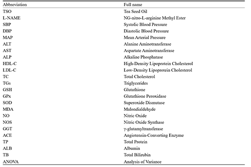
List of abbreviations |
Availability of data and materials
The datasets used and/or analyzed during the current study are available from the corresponding author on resonable rquest.
Competing interests
The authors declare no conflict of interest.
Ethics approval and consent to participate
Authors declared that all experiments were carried out with strict compliance to the “Principle of Laboratory Animal Care” and ethical guidelines for investigation of experimental pain in conscious animals. The project was approved by the University of Ibadan Animal Care and Use Research Ethics Committee.
Funding
This project was funded by the authors and the management of Cocoa Research Institute of Nigeria (CRIN).
Authors’ Contributions
LEY: Conceptualization, supervision and final correction of draft. JFA and AAO: Methodology, data collection, writing-original draft preparation. LEY and JFA: Editing. All authors have read and approved the final manuscript.
Adedapo A.D.A., Ajayi A.M., Ekwunife N.L., Falayi O.O., Oyagbemi A., Omobowale T.O., and Adedapo A.A., 2020, Antihypertensive effect of Phragmanthera incana (Schum) balle on NG-nitro-L-Arginine methyl ester (L-NAME) induced hypertensive rats, Journal of Ethnopharmacology, 257: 112888.
https://doi.org/10.1016/j.jep.2020.112888
Akindele M.O., 2014, An intervention programme for management of overweight and obese nigerians in lagos state, Nigeria.
Babson A.L., Greeley S.J., Coleman C.M., and Philips G.D., 1966, Phenolphthalein monophosphate as a substrate for serum alkaline phosphatase, Clinical Chemistry, 12(8): 482-490.
https://doi.org/10.1093/clinchem/12.8.482
Banerjee B.D., Seth V., Bhattacharya A., Pasha S.T., and Chakraborty A.K., 1999, Biochemical effects of some pesticides on lipid peroxidation and free-radical scavengers, Toxicology Letters, 107(1-3): 33-47.
https://doi.org/10.1016/S0378-4274(99)00029-6
Biggs H.G., Erikson J.M., and Moorehead W.R., 1975, A manual colorimetric assay of triglycerides in serum, Clinical Chemistry, 21(3): 437-441.
https://doi.org/10.1093/clinchem/21.3.437
Ben-Aicha S., Badimon L., and Vilahur G., 2020, Advances in HDL: much more than lipid transporters, International Journal of Molecular Sciences, 21(3): 732.
https://doi.org/10.3390/ijms21030732
Doumas B.T., Watson W.A., and Biggs H.G., 1971, Albumin standards and the measurement of serum albumin with bromcresol green, Clinica Chimica Acta, 31(1): 87-96.
https://doi.org/10.1016/0009-8981(71)90365-2
Fuchs D., De Graaf Y., Van Kerckhoven R., and Draijer R., 2014, Effect of tea theaflavins and catechins on microvascular function, Nutrients, 6(12): 5772-5785.
https://doi.org/10.3390/nu6125772
Goth L., 1991, A simple method for determination of serum catalase activity and revision of reference range, Clinica Chimica Acta, 196(2-3): 143-151.
https://doi.org/10.1016/0009-8981(91)90067-M
Guzik T.J., and Touyz R.M., 2017, Oxidative Stress, Inflammation, and vascular aging in hypertension, Hypertension, 70(4): 660-667.
https://doi.org/10.1161/HYPERTENSIONAHA.117.07802
Irving G.W., 1958, AOCS Commentary: guidelines for research on fats and oils in the united states department of agriculture, Journal of the American Oil Chemists' Society, 35(4): 168-170.
https://doi.org/10.1007/BF02640600
Krasylenko Y.A., Yemets A.I., and Blume Y.B., 2019, Nitric oxide synthase inhibitor L-NAME affects Arabidopsis root growth, morphology, and microtubule organization, Cell Biology International, 43(9): 1049-1055.
https://doi.org/10.1002/cbin.10880
Khan N., and Mukhtar H., 2018, Tea polyphenols in promotion of human health, Nutrients, 11(1): 39.
https://doi.org/10.3390/nu11010039
Khatana C., Saini N.K., Chakrabarti S., Saini V., Sharma A., Saini R.V., and Saini A.K., 2020, Mechanistic Insights into the oxidized low-density lipoprotein-induced atherosclerosis, Oxidative Medicine and Cellular Longevity, 2020(1): 5245308.
https://doi.org/10.1155/2020/5245308
Kibret K.T., and Mesfin Y.M., 2015, Prevalence of hypertension in ethiopia: a systematic meta-analysis, Public Health Reviews, 36(1): 14.
https://doi.org/10.1186/s40985-015-0014-z
Kinoshita C., Saze K.I., Kumata S., Matsuki T., and Homma S., 1996, A simplified method for the estimation of glutathione peroxidase activity and selenium concentration in bovine blood, Journal of Dairy Science, 79(9): 1543-1548.
https://doi.org/10.3168/jds.S0022-0302(96)76515-3
Krasylenko Y.A., Yemets A.I., and Blume Y.B., 2019, Nitric oxide synthase inhibitor l-name affects arabidopsis root growth, morphology, and microtubule organization, Cell Biology International, 43(9): 1049-1055.
https://doi.org/10.1002/cbin.10880
Krishnaveni P., and Gowda V.M., 2015, Assessing the validity of friedewald's formula and anandraja's formula for serum LDL-cholesterol calculation, Journal of Clinical and Diagnostic Research, 9(12): BC01-BC04.
https://doi.org/10.7860/JCDR/2015/16850.6870
Lee Y., and Siddiqui W.J., 2019, Cholesterol Levels, StatPearls Publishing, Treasure Island, FL, USA, pp.1-23.
Li B., He X., Lei S.S., Zhou F.C., Zhang N.Y., Chen Y.H., Wang Y.Z., Su J., Yu J.J., Li L.Z., and Zheng X., 2020, Hypertensive rats treated chronically with Nω-Nitro-L-Arginine methyl ester (L-NAME) induced disorder of hepatic fatty acid metabolism and intestinal pathophysiology, Frontiers in Pharmacology, 10: 1677.
https://doi.org/10.3389/fphar.2019.01677
Linné J.J., and Ringsrud K.M., eds., 1979, Basic Techniques for the Medical Laboratory, 2nd ed., McGraw-Hill, New York, USA, pp.1-523.
Mahmoudi R., Azizi A., Abedini S., Jahromi V.H., Abidi H., and Barmak M.J., 2018, Green tea improves rat sperm quality and reduces cadmium chloride damage effect in spermatogenesis cycle, Journal of Medicine and Life, 11(4): 371-375.
https://doi.org/10.25122/jml-2018-0005
Malloy H.T., and Evelyn K.A., 1937, The determination of bilirubin with the photoelectric colorimeter, Journal of Biological Chemistry, 119(2): 481-490.
https://doi.org/10.1016/S0021-9258(18)74392-5
Marklund S., and Marklund G., 1974, Involvement of the superoxide anion radical in the autoxidation of pyrogallol and a convenient assay for superoxide dismutase, European Journal of Biochemistry, 47(3): 469-474.
https://doi.org/10.1111/j.1432-1033.1974.tb03714.x
Maurya R., Namdeo M., 2021, Superoxide dismutase: a key enzyme for the survival of intracellular pathogens in host, Reactive Oxygen Species.
https://doi.org/10.5772/intechopen.100322
Metchi Donfack M.F., Atsamo A.D., Temdié Guemmogne R.J., Ngouateu Kenfack O.B., Dongmo A.B., and Dimo T., 2021, Antihypertensive effects of the Vitex cienkowskii (Verbenaceae) stem-bark extract on L-NAME-induced hypertensive rats, Evidence-Based Complementary and Alternative Medicine, 2021: 6668919.
https://doi.org/10.5772/intechopen.100322
Muntner P., Shimbo D., Carey R.M., Charleston J.B., Gaillard T., Misra S., and Wright J.T. Jr., 2019, Measurement of blood pressure in humans: a scientific statement from the american heart association, Hypertension, 73(5): e35-e66.
https://doi.org/10.1161/HYP.0000000000000087
Musial C., Kuban-Jankowska A., and Gorska-Ponikowska M., 2020, Beneficial properties of green tea catechins, International Journal of Molecular Sciences, 21(5): 1744.
https://doi.org/10.3390/ijms21051744
Nwanjo H.U., and Alumanah E.O., 2006, Effect of aqueous extract of gongronema latifolium on some indices of liver function in rats, Global Journal of Medical Sciences, 5(1): 17-20.
https://doi.org/10.4314/gjms.v5i1.10142
Oliveira G.M.M.D., Brant L.C.C., Polanczyk C.A., Malta D.C., Biolo A., Nascimento B.R., and Ribeiro A.L.P., 2022, Cardiovascular statistics-Brazil 2021, Arquivos Brasileiros de Cardiologia, 118: 115-373.
https://doi.org/10.36660/abc.20211012
Okhawa H., Ohishi N., and Yagi K., 1979, Assay for lipid peroxides in animal tissues by thiobarbituric acid reaction, Analytical Biochemistry, 95(2): 351-358.
https://doi.org/10.1016/0003-2697(79)90738-3
Packard C.J., Boren J., and Taskinen M.R., 2020, Causes and consequences of hypertriglyceridemia, Frontiers in Endocrinology, 11: 252.
https://doi.org/10.3389/fendo.2020.00252
Panday S., Talreja R., and Kavdia M., 2020, The role of glutathione and glutathione peroxidase in regulating cellular levels of reactive oxygen and nitrogen species, Microvascular Research, 131: 104019.
https://doi.org/10.1016/j.mvr.2020.104010
Rahman S., Islam S., Haque T., Kathak R.R., and Ali N., 2020, Association between serum liver enzymes and hypertension: a cross-sectional study in bangladeshi adults, BMC Cardiovascular Disorders, 20: 1-7.
https://doi.org/10.1186/s12872-020-01411-6
Reitman S., and Frankel S., 1957, A colorimetric determination of serum glutamic oxaloacetic and glutamic pyruvic transaminase, American Journal of Clinical Pathology, 28(1): 56-63.
https://doi.org/10.1093/ajcp/28.1.56
Roeschlau P., Bernt E., and Gruber W., 1974, Enzymatic determination of total cholesterol in serum, Zeitschrift für Klinische Chemie und Klinische Biochemie, 12: 226-227.
Samavat H., Newman A.R., Wang R., Yuan J.M., Wu A.H., and Kurzer M.S., 2016, Effects of green tea catechin extract on serum lipids in postmenopausal women: a randomized, placebo-controlled clinical trial, American Journal of Clinical Nutrition, 103(6): 1671-1682.
https://doi.org/10.3945/ajcn.116.137075
Suttisansanee U., Kunkeaw T., Thatsanasuwan N., Tonglim J., and Temviriyanukul P., 2019, The investigation on cholinesterases and BACE1 inhibitory activities in various tea infusions, Walailak Journal of Science and Technology, 16(3): 165-174.
https://doi.org/10.48048/wjst.2019.6221
Tan B.L., Norhaizan M.E., Liew W.P.P., and Sulaiman R.H., 2018, Antioxidant and oxidative stress: a mutual interplay in age-related diseases, Frontiers in Pharmacology, 9: 1162.
https://doi.org/10.3389/fphar.2018.01162
Tawfeek H.M., Faisal W., and Soliman G.M., 2018, Enalapril maleate orally disintegrating tablets: tableting and in vivo evaluation in hypertensive rats, Pharmaceutical Development and Technology, 23(5): 496-503.
https://doi.org/10.1080/10837450.2017.1329318
Vitolo M., 2021, Decomposition of hydrogen peroxide by catalase, World Journal of Pharmacy and Pharmaceutical Sciences, 10(8): 47-56.
Wang C., Han J., Pu Y., and Wang X., 2022, Tea (Camellia sinensis): A Review of Nutritional Composition, Potential Applications, and Omics Research, Applied Sciences, 12(12): 5874.
https://doi.org/10.3390/app12125874
Wang J.G., Zhang W., Li Y., and Liu L., 2023, Hypertension in china: epidemiology and treatment initiatives, Nature Reviews Cardiology, 20: 1-15.
https://doi.org/10.1038/s41569-022-00829-z
Wang S.T., Cui W.Q., Pan D., Jiang M., Chang B., and Sang L.X., 2020, Tea polyphenols and their chemopreventive and therapeutic effects on colorectal cancer, World Journal of Gastroenterology, 26(6): 562-575.
Warnick G.R., Nauck M., and Rifai N., 2001, Evolution of Methods for Measurement of HDL-cholesterol: from ultracentrifugation to homogeneous assays, Clinical Chemistry, 47(9): 1579-1596.
https://doi.org/10.1093/clinchem/47.9.1579
Yan Z., Zhong Y., Duan Y., Chen Q., and Li F., 2020, Antioxidant mechanism of tea polyphenols and its impact on health benefits, Animal Nutrition, 6(2): 115-123.
https://doi.org/10.1016/j.aninu.2020.01.001
Zhao Y., Vanhoutte P.M., and Leung S.W., 2015, Vascular nitric oxide: beyond eNOS, Journal of Pharmacological Sciences, 129(2): 83-94.
https://doi.org/10.1016/j.jphs.2015.09.002


. FPDF(win)
. FPDF(mac)
. HTML
. Online fPDF
Associated material
. Readers' comments
Other articles by authors
. L. E Yahaya
. J. F. Atanda
. A. A. Oyagbemi
Related articles
. Hypertension
. Blood Pressure
. Tea seed oil
. Oxidative stress
. Antioxidant enzymes
Tools
. Post a comment